5K6P

The NMR structure of the m domain tri-helix bundle and C2 of human cardiac Myosin Binding Protein C


Experimental Data Snapshot

  • Method: SOLUTION NMR
  • Conformers Calculated: 500 
  • Conformers Submitted: 20 
  • Selection Criteria: structures with the lowest energy 

wwPDB Validation   3D Report Full Report


This is version 1.4 of the entry. See complete history


Literature

A Highly Conserved Yet Flexible Linker Is Part of a Polymorphic Protein-Binding Domain in Myosin-Binding Protein C.

Michie, K.A.Kwan, A.H.Tung, C.S.Guss, J.M.Trewhella, J.

(2016) Structure 24: 2000-2007

  • DOI: https://doi.org/10.1016/j.str.2016.08.018
  • Primary Citation of Related Structures:  
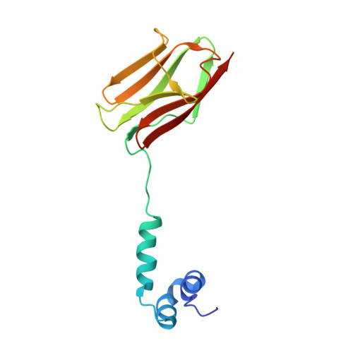
    5K6P

  • PubMed Abstract: 

    The nuclear magnetic resonance (NMR) structure of the tri-helix bundle (THB) of the m-domain plus C2 (ΔmC2) of myosin-binding protein C (MyBP-C) has revealed a highly flexible seven-residue linker between the structured THB and C2. Bioinformatics shows significant patterns of conservation across the THB-linker sequence, with the linker containing a strictly conserved serine in all MyBP-C isoforms. Clinically linked mutations further support the functional significance of the THB-linker region. NMR, small-angle X-ray scattering, and binding studies show the THB-linker plus the first ten residues of C2 undergo dramatic changes when ΔmC2 binds Ca 2+ -calmodulin, with the linker and C2 N-terminal residues contributing significantly to the affinity. Modeling of all available experimental data indicates that the THB tertiary structure must be disrupted to form the complex. These results are discussed in the context of the THB-linker and the N-terminal residues of C2 forming a polymorphic binding domain that could accommodate multiple binding partners in the dynamic sarcomere.


  • Organizational Affiliation

    School of Life and Environmental Sciences, The University of Sydney, NSW 2006, Australia.


Macromolecules
Find similar proteins by:  (by identity cutoff)  |  3D Structure
Entity ID: 1
MoleculeChains Sequence LengthOrganismDetailsImage
Myosin-binding protein C, cardiac-type137Homo sapiensMutation(s): 0 
Gene Names: MYBPC3
UniProt & NIH Common Fund Data Resources
Find proteins for Q14896 (Homo sapiens)
Explore Q14896 
Go to UniProtKB:  Q14896
PHAROS:  Q14896
GTEx:  ENSG00000134571 
Entity Groups  
Sequence Clusters30% Identity50% Identity70% Identity90% Identity95% Identity100% Identity
UniProt GroupQ14896
Sequence Annotations
Expand
  • Reference Sequence
Experimental Data & Validation

Experimental Data

  • Method: SOLUTION NMR
  • Conformers Calculated: 500 
  • Conformers Submitted: 20 
  • Selection Criteria: structures with the lowest energy 

Structure Validation

View Full Validation Report


Entry History & Funding Information

Deposition Data


Funding OrganizationLocationGrant Number
Australian Research Council (ARC)AustraliaDP120103841

Revision History  (Full details and data files)

  • Version 1.0: 2016-11-09
    Type: Initial release
  • Version 1.1: 2016-11-16
    Changes: Database references
  • Version 1.2: 2017-11-01
    Changes: Author supporting evidence, Data collection, Database references, Structure summary
  • Version 1.3: 2020-01-01
    Changes: Author supporting evidence, Data collection
  • Version 1.4: 2023-06-14
    Changes: Database references, Other