5K48

VIM-2 Metallo Beta Lactamase in complex with 3-(mercaptomethyl)-[1,1'-biphenyl]-4-carboxylic acid


Experimental Data Snapshot

  • Method: X-RAY DIFFRACTION
  • Resolution: 1.74 Å
  • R-Value Free: 0.194 
  • R-Value Work: 0.166 
  • R-Value Observed: 0.167 

wwPDB Validation   3D Report Full Report


Ligand Structure Quality Assessment 


This is version 1.3 of the entry. See complete history


Literature

In Silico Fragment-Based Design Identifies Subfamily B1 Metallo-beta-lactamase Inhibitors.

Cain, R.Brem, J.Zollman, D.McDonough, M.A.Johnson, R.M.Spencer, J.Makena, A.Abboud, M.I.Cahill, S.Lee, S.Y.McHugh, P.J.Schofield, C.J.Fishwick, C.W.G.

(2018) J Med Chem 61: 1255-1260

  • DOI: https://doi.org/10.1021/acs.jmedchem.7b01728
  • Primary Citation of Related Structures:  
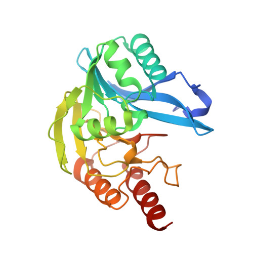
    5K48

  • PubMed Abstract: 

    Zinc ion-dependent β-lactamases (MBLs) catalyze the hydrolysis of almost all β-lactam antibiotics and resist the action of clinically available β-lactamase inhibitors. We report how application of in silico fragment-based molecular design employing thiol-mediated metal anchorage leads to potent MBL inhibitors. The new inhibitors manifest potent inhibition of clinically important B1 subfamily MBLs, including the widespread NDM-1, IMP-1, and VIM-2 enzymes; with lower potency, some of them also inhibit clinically relevant Class A and D serine-β-lactamases. The inhibitors show selectivity for bacterial MBL enzymes compared to that for human MBL fold nucleases. Cocrystallization of one inhibitor, which shows potentiation of Meropenem activity against MBL-expressing Enterobacteriaceae, with VIM-2 reveals an unexpected binding mode, involving interactions with residues from conserved active site bordering loops.


  • Organizational Affiliation

    School of Chemistry, University of Leeds , Leeds LS2 9JT, United Kingdom.


Macromolecules
Find similar proteins by:  (by identity cutoff)  |  3D Structure
Entity ID: 1
MoleculeChains Sequence LengthOrganismDetailsImage
Beta-lactamase VIM-2
A, B
242Pseudomonas aeruginosaMutation(s): 0 
Gene Names: blaVIM-2bla vim-2bla-VIM-2blasVIM-2blaVIM2blmVIM-2PAERUG_P32_London_17_VIM_2_10_11_06255
UniProt
Find proteins for Q9K2N0 (Pseudomonas aeruginosa)
Explore Q9K2N0 
Go to UniProtKB:  Q9K2N0
Entity Groups  
Sequence Clusters30% Identity50% Identity70% Identity90% Identity95% Identity100% Identity
UniProt GroupQ9K2N0
Sequence Annotations
Expand
  • Reference Sequence
Small Molecules
Ligands 3 Unique
IDChains Name / Formula / InChI Key2D Diagram3D Interactions
S5Z
Query on S5Z

Download Ideal Coordinates CCD File 
C [auth A],
J [auth B]
4-phenyl-2-(sulfanylmethyl)benzoic acid
C14 H12 O2 S
OJGNJYVRHUYKQS-UHFFFAOYSA-N
ZN
Query on ZN

Download Ideal Coordinates CCD File 
G [auth A]
H [auth A]
I [auth A]
M [auth B]
N [auth B]
G [auth A],
H [auth A],
I [auth A],
M [auth B],
N [auth B],
O [auth B]
ZINC ION
Zn
PTFCDOFLOPIGGS-UHFFFAOYSA-N
FMT
Query on FMT

Download Ideal Coordinates CCD File 
D [auth A],
E [auth A],
F [auth A],
K [auth B],
L [auth B]
FORMIC ACID
C H2 O2
BDAGIHXWWSANSR-UHFFFAOYSA-N
Experimental Data & Validation

Experimental Data

  • Method: X-RAY DIFFRACTION
  • Resolution: 1.74 Å
  • R-Value Free: 0.194 
  • R-Value Work: 0.166 
  • R-Value Observed: 0.167 
  • Space Group: C 1 2 1
Unit Cell:
Length ( Å )Angle ( ˚ )
a = 102.888α = 90
b = 79.542β = 130.6
c = 67.892γ = 90
Software Package:
Software NamePurpose
PHENIXrefinement
HKL-2000data reduction
HKL-2000data scaling
PHASERphasing

Structure Validation

View Full Validation Report


Ligand Structure Quality Assessment 


Entry History 

Deposition Data

Revision History  (Full details and data files)

  • Version 1.0: 2017-06-07
    Type: Initial release
  • Version 1.1: 2018-01-17
    Changes: Database references
  • Version 1.2: 2018-02-21
    Changes: Database references
  • Version 1.3: 2024-01-10
    Changes: Data collection, Database references, Derived calculations, Refinement description