Insights into an efficient light-driven hybrid P450 BM3 enzyme from crystallographic, spectroscopic and biochemical studies.
Spradlin, J., Lee, D., Mahadevan, S., Mahomed, M., Tang, L., Lam, Q., Colbert, A., Shafaat, O.S., Goodin, D., Kloos, M., Kato, M., Cheruzel, L.E.(2016) Biochim Biophys Acta 1864: 1732-1738
- PubMed: 27639964
- DOI: https://doi.org/10.1016/j.bbapap.2016.09.005
- Primary Citation of Related Structures:
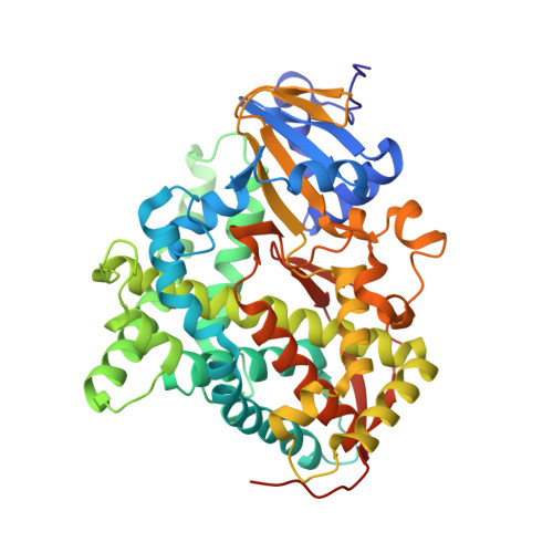
5JQ2, 5JTD - PubMed Abstract:
In order to perform selective CH functionalization upon visible light irradiation, Ru(II)-diimine functionalized P450 heme enzymes have been developed. The sL407C-1 enzyme containing the Ru(bpy) 2 PhenA (bpy=2,2'-bipyridine and PhenA=5-acetamido-1,10-phenanthroline) photosensitizer (1) covalently attached to the non-native single cysteine L407C of the P450BM3 heme domain mutant, displays high photocatalytic activity in the selective CH bond hydroxylation of several substrates. A combination of X-ray crystallography, site-directed mutagenesis, transient absorption measurements and enzymatic assays was used to gain insights into its photocatalytic activity and electron transfer pathway. The crystal structure of the sL407C-1 enzyme was solved in the open and closed conformations revealing a through-space electron transfer pathway involving highly conserved, F393 and Q403, residues. Several mutations of these residues (F393A, F393W or Q403W) were introduced to probe their roles in the overall reaction. Transient absorption measurements confirm rapid electron transfer as heme reduction is observed in all four hybrid enzymes. Compared to the parent sL407C-1, photocatalytic activity was negligible in the dF393A-1 enzyme while 60% increase in activity with total turnover numbers of 420 and 90% product conversion was observed with the dQ403W-1 mutant. In the sL407C-1 enzyme, the photosensitizer is ideally located to rapidly deliver electrons, using the naturally occurring electron transfer pathway, to the heme center in order to activate molecular dioxygen and sustain photocatalytic activity. The results shed light on the design of efficient light-driven biocatalysts and the approach can be generalized to other members of the P450 superfamily.
Organizational Affiliation:
San José State University, Department of Chemistry, One Washington Square, San José, CA, United States.