5JG8

Crystal structure of human acid sphingomyelinase


Experimental Data Snapshot

  • Method: X-RAY DIFFRACTION
  • Resolution: 2.80 Å
  • R-Value Free: 0.279 
  • R-Value Work: 0.230 
  • R-Value Observed: 0.232 

wwPDB Validation   3D Report Full Report


Ligand Structure Quality Assessment 


This is version 2.1 of the entry. See complete history


Literature

Structure of Human Acid Sphingomyelinase Reveals the Role of the Saposin Domain in Activating Substrate Hydrolysis.

Xiong, Z.J.Huang, J.Poda, G.Pomes, R.Prive, G.G.

(2016) J Mol Biol 428: 3026-3042

  • DOI: https://doi.org/10.1016/j.jmb.2016.06.012
  • Primary Citation of Related Structures:  
    5JG8

  • PubMed Abstract: 

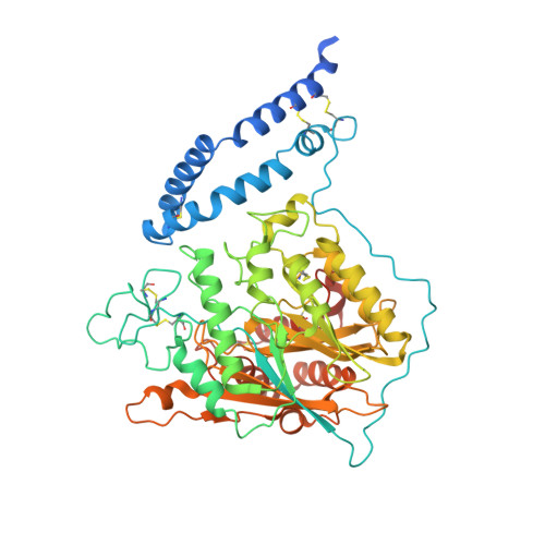
    Acid sphingomyelinase (ASM) is a lysosomal phosphodiesterase that catalyzes the hydrolysis of sphingomyelin to produce ceramide and phosphocholine. While other lysosomal sphingolipid hydrolases require a saposin activator protein for full activity, the ASM polypeptide incorporates a built-in N-terminal saposin domain and does not require an external activator protein. Here, we report the crystal structure of human ASM and describe the organization of the three main regions of the enzyme: the N-terminal saposin domain, the proline-rich connector, and the catalytic domain. The saposin domain is tightly associated along an edge of the large, bowl-shaped catalytic domain and adopts an open form that exposes a hydrophobic concave surface approximately 30Å from the catalytic center. The calculated electrostatic potential of the enzyme is electropositive at the acidic pH of the lysosome, consistent with the strict requirement for the presence of acidic lipids in target membranes. Docking studies indicate that sphingomyelin binds with the ceramide-phosphate group positioned at the binuclear zinc center and molecular dynamic simulations indicate that the intrinsic flexibility of the saposin domain is important for monomer-dimer exchange and for membrane interactions. Overall, ASM uses a combination of electrostatic and hydrophobic interactions to cause local disruptions of target bilayers in order to bring the lipid headgroup to the catalytic center in a membrane-bound reaction.


  • Organizational Affiliation

    Department of Biochemistry, University of Toronto, Toronto, Ontario, Canada.


Macromolecules
Find similar proteins by:  (by identity cutoff)  |  3D Structure
Entity ID: 1
MoleculeChains Sequence LengthOrganismDetailsImage
Sphingomyelin phosphodiesterase
A, B
586Homo sapiensMutation(s): 1 
Gene Names: SMPD1ASM
EC: 3.1.4.12
UniProt & NIH Common Fund Data Resources
Find proteins for P17405 (Homo sapiens)
Explore P17405 
Go to UniProtKB:  P17405
PHAROS:  P17405
GTEx:  ENSG00000166311 
Entity Groups  
Sequence Clusters30% Identity50% Identity70% Identity90% Identity95% Identity100% Identity
UniProt GroupP17405
Sequence Annotations
Expand
  • Reference Sequence
Oligosaccharides

Help

Entity ID: 2
MoleculeChains Length2D Diagram Glycosylation3D Interactions
2-acetamido-2-deoxy-beta-D-glucopyranose-(1-4)-2-acetamido-2-deoxy-beta-D-glucopyranose
C, D, H, I
2N-Glycosylation
Glycosylation Resources
GlyTouCan:  G42666HT
GlyCosmos:  G42666HT
GlyGen:  G42666HT
Entity ID: 3
MoleculeChains Length2D Diagram Glycosylation3D Interactions
2-acetamido-2-deoxy-beta-D-glucopyranose-(1-3)-2-acetamido-2-deoxy-beta-D-glucopyranose
E
2N-Glycosylation
Glycosylation Resources
GlyTouCan:  G79924RM
GlyCosmos:  G79924RM
GlyGen:  G79924RM
Entity ID: 4
MoleculeChains Length2D Diagram Glycosylation3D Interactions
alpha-D-mannopyranose-(1-4)-2-acetamido-2-deoxy-beta-D-glucopyranose-(1-4)-2-acetamido-2-deoxy-beta-D-glucopyranose
F, G
3N-Glycosylation
Glycosylation Resources
GlyTouCan:  G62182OO
GlyCosmos:  G62182OO
GlyGen:  G62182OO
Small Molecules
Ligands 3 Unique
IDChains Name / Formula / InChI Key2D Diagram3D Interactions
NAG
Query on NAG

Download Ideal Coordinates CCD File 
M [auth A],
N [auth A],
O [auth A],
S [auth B],
T [auth B]
2-acetamido-2-deoxy-beta-D-glucopyranose
C8 H15 N O6
OVRNDRQMDRJTHS-FMDGEEDCSA-N
ZN
Query on ZN

Download Ideal Coordinates CCD File 
J [auth A],
K [auth A],
P [auth B],
Q [auth B]
ZINC ION
Zn
PTFCDOFLOPIGGS-UHFFFAOYSA-N
ACT
Query on ACT

Download Ideal Coordinates CCD File 
L [auth A],
R [auth B]
ACETATE ION
C2 H3 O2
QTBSBXVTEAMEQO-UHFFFAOYSA-M
Experimental Data & Validation

Experimental Data

  • Method: X-RAY DIFFRACTION
  • Resolution: 2.80 Å
  • R-Value Free: 0.279 
  • R-Value Work: 0.230 
  • R-Value Observed: 0.232 
  • Space Group: P 21 21 21
Unit Cell:
Length ( Å )Angle ( ˚ )
a = 69.531α = 90
b = 143.658β = 90
c = 193.62γ = 90
Software Package:
Software NamePurpose
PHENIXrefinement
HKL-2000data reduction
Aimlessdata scaling
PHENIXphasing
PDB_EXTRACTdata extraction

Structure Validation

View Full Validation Report


Ligand Structure Quality Assessment 


Entry History 

Deposition Data

Revision History  (Full details and data files)

  • Version 1.0: 2016-07-06
    Type: Initial release
  • Version 1.1: 2016-07-13
    Changes: Database references
  • Version 1.2: 2016-08-17
    Changes: Database references
  • Version 2.0: 2020-07-29
    Type: Remediation
    Reason: Carbohydrate remediation
    Changes: Advisory, Atomic model, Data collection, Database references, Derived calculations, Structure summary
  • Version 2.1: 2023-09-27
    Changes: Data collection, Database references, Refinement description, Structure summary