5J76

Structure of Lectin from Colocasia esculenta(L.) Schott


Experimental Data Snapshot

  • Method: X-RAY DIFFRACTION
  • Resolution: 2.10 Å
  • R-Value Free: 0.204 
  • R-Value Work: 0.165 
  • R-Value Observed: 0.167 

wwPDB Validation   3D Report Full Report


This is version 1.2 of the entry. See complete history


Literature

Structural analysis of beta-prism lectin from Colocasia esculenta (L.) S chott.

Vajravijayan, S.Pletnev, S.Pletnev, V.Z.Nandhagopal, N.Gunasekaran, K.

(2016) Int J Biol Macromol 91: 518-523

  • DOI: https://doi.org/10.1016/j.ijbiomac.2016.05.048
  • Primary Citation of Related Structures:  
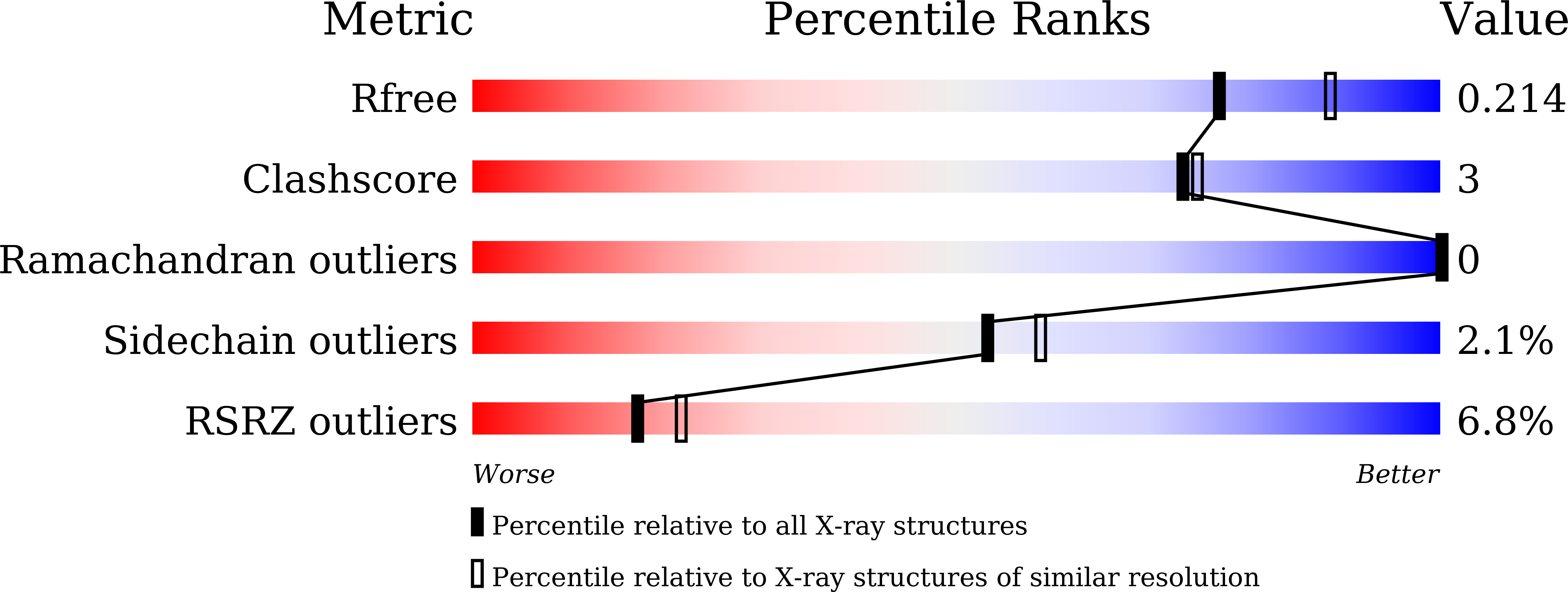
    5J76

  • PubMed Abstract: 

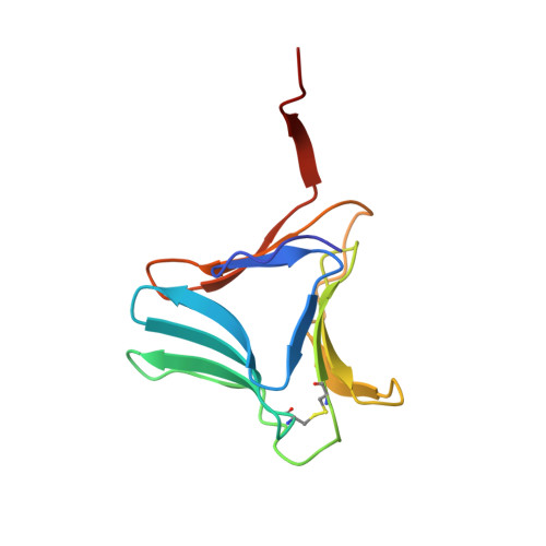
    The Mannose-binding β-Prism Colocasia esculenta lectin (β-PCL) was purified from tubers using ion exchange chromatography. The purified β-PCL appeared as a single band of ∼12kDa on SDS-PAGE. β-PCL crystallizes in trigonal space group P3121 and diffracted to a resolution of 2.1Å. The structure was solved using Molecular replacement using Crocus vernus lectin (PDB: 3MEZ) as a model. From the final refined model to an R-factor of 16.5% and an Rfree of 20.4%, it has been observed that the biological unit consists of two β-Prism domains augmented through C-terminals swap over to form one of faces for each domain. Cα superposition of individual domains of β-PCL with individual domains of other related structures and superposition of whole protein structures were carried out. The higher RMS deviation for the superposition of whole structures suggest that β-prism domains assume different orientation in each structure.


  • Organizational Affiliation

    Centre of Advanced Study in Crystallography and Biophysics, University of Madras, Guindy Campus, Chennai 600 025, India.


Macromolecules
Find similar proteins by:  (by identity cutoff)  |  3D Structure
Entity ID: 1
MoleculeChains Sequence LengthOrganismDetailsImage
12kD storage protein109Colocasia esculentaMutation(s): 0 
UniProt
Find proteins for Q39487 (Colocasia esculenta)
Explore Q39487 
Go to UniProtKB:  Q39487
Entity Groups  
Sequence Clusters30% Identity50% Identity70% Identity90% Identity95% Identity100% Identity
UniProt GroupQ39487
Sequence Annotations
Expand
  • Reference Sequence
Find similar proteins by:  (by identity cutoff)  |  3D Structure
Entity ID: 2
MoleculeChains Sequence LengthOrganismDetailsImage
12kD storage protein111Colocasia esculentaMutation(s): 0 
UniProt
Find proteins for Q39487 (Colocasia esculenta)
Explore Q39487 
Go to UniProtKB:  Q39487
Entity Groups  
Sequence Clusters30% Identity50% Identity70% Identity90% Identity95% Identity100% Identity
UniProt GroupQ39487
Sequence Annotations
Expand
  • Reference Sequence
Experimental Data & Validation

Experimental Data

  • Method: X-RAY DIFFRACTION
  • Resolution: 2.10 Å
  • R-Value Free: 0.204 
  • R-Value Work: 0.165 
  • R-Value Observed: 0.167 
  • Space Group: P 31 2 1
Unit Cell:
Length ( Å )Angle ( ˚ )
a = 75.471α = 90
b = 75.471β = 90
c = 121.177γ = 120
Software Package:
Software NamePurpose
HKL-2000data collection
SCALEPACKdata scaling
MOLREPphasing
REFMACrefinement
PDB_EXTRACTdata extraction
HKL-2000data reduction
MOLREPphasing

Structure Validation

View Full Validation Report


Entry History 

Deposition Data

Revision History  (Full details and data files)

  • Version 1.0: 2016-06-22
    Type: Initial release
  • Version 1.1: 2016-06-29
    Changes: Database references
  • Version 1.2: 2023-11-08
    Changes: Data collection, Database references, Derived calculations, Refinement description