5IT5

Thermus thermophilus PilB core ATPase region


Experimental Data Snapshot

  • Method: X-RAY DIFFRACTION
  • Resolution: 2.65 Å
  • R-Value Free: 0.274 
  • R-Value Work: 0.208 
  • R-Value Observed: 0.210 

wwPDB Validation   3D Report Full Report


Ligand Structure Quality Assessment 


This is version 1.5 of the entry. See complete history


Literature

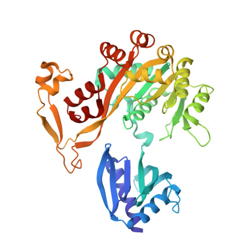
Crystal Structure of a Type IV Pilus Assembly ATPase: Insights into the Molecular Mechanism of PilB from Thermus thermophilus.

Mancl, J.M.Black, W.P.Robinson, H.Yang, Z.Schubot, F.D.

(2016) Structure 24: 1886-1897

  • DOI: https://doi.org/10.1016/j.str.2016.08.010
  • Primary Citation of Related Structures:  
    5IT5

  • PubMed Abstract: 

    Type IV pili (T4P) mediate bacterial motility and virulence. The PilB/GspE family ATPases power the assembly of T4P and type 2 secretion systems. We determined the structure of the ATPase region of PilB (PilB ATP ) in complex with ATPγS to provide a model of a T4P assembly ATPase and a view of a PilB/GspE family hexamer at better than 3-Å resolution. Spatial positioning and conformations of the protomers suggest a mechanism of force generation. All six PilB ATP protomers contain bound ATPγS. Two protomers form a closed conformation poised for ATP hydrolysis. The other four molecules assume an open conformation but separate into two pairs with distinct active-site accessibilities. We propose that one pair represents the post-hydrolysis phase while the other pair appears poised for ADP/ATP exchange. Collectively, the data suggest that T4P assembly is powered by coordinating concurrent substrate binding with ATP hydrolysis across the PilB hexamer.


  • Organizational Affiliation

    Department of Biological Sciences, Virginia Polytechnic Institute and State University, 125 Life Sciences 1 (MC 0910), 970 Washington Street Southwest, Blacksburg, VA 24061, USA.


Macromolecules
Find similar proteins by:  (by identity cutoff)  |  3D Structure
Entity ID: 1
MoleculeChains Sequence LengthOrganismDetailsImage
ATP binding motif-containing protein PilF385Thermus thermophilusMutation(s): 0 
Gene Names: pilF
UniProt
Find proteins for Q5SLC9 (Thermus thermophilus (strain ATCC 27634 / DSM 579 / HB8))
Explore Q5SLC9 
Go to UniProtKB:  Q5SLC9
Entity Groups  
Sequence Clusters30% Identity50% Identity70% Identity90% Identity95% Identity100% Identity
UniProt GroupQ5SLC9
Sequence Annotations
Expand
  • Reference Sequence
Small Molecules
Ligands 4 Unique
IDChains Name / Formula / InChI Key2D Diagram3D Interactions
AGS
Query on AGS

Download Ideal Coordinates CCD File 
I [auth A],
K [auth B],
N [auth E],
Q [auth C],
S [auth D]
PHOSPHOTHIOPHOSPHORIC ACID-ADENYLATE ESTER
C10 H16 N5 O12 P3 S
NLTUCYMLOPLUHL-KQYNXXCUSA-N
ATP
Query on ATP

Download Ideal Coordinates CCD File 
H [auth F]ADENOSINE-5'-TRIPHOSPHATE
C10 H16 N5 O13 P3
ZKHQWZAMYRWXGA-KQYNXXCUSA-N
ZN
Query on ZN

Download Ideal Coordinates CCD File 
G [auth F]
J [auth A]
L [auth B]
O [auth E]
R [auth C]
G [auth F],
J [auth A],
L [auth B],
O [auth E],
R [auth C],
T [auth D]
ZINC ION
Zn
PTFCDOFLOPIGGS-UHFFFAOYSA-N
MG
Query on MG

Download Ideal Coordinates CCD File 
M [auth B],
P [auth E]
MAGNESIUM ION
Mg
JLVVSXFLKOJNIY-UHFFFAOYSA-N
Experimental Data & Validation

Experimental Data

  • Method: X-RAY DIFFRACTION
  • Resolution: 2.65 Å
  • R-Value Free: 0.274 
  • R-Value Work: 0.208 
  • R-Value Observed: 0.210 
  • Space Group: P 2 21 21
Unit Cell:
Length ( Å )Angle ( ˚ )
a = 107.55α = 90
b = 133.56β = 90
c = 208.42γ = 90
Software Package:
Software NamePurpose
PHENIXrefinement
iMOSFLMdata reduction
SCALAdata scaling
PHASERphasing

Structure Validation

View Full Validation Report


Ligand Structure Quality Assessment 


Entry History & Funding Information

Deposition Data


Funding OrganizationLocationGrant Number
National Science Foundation (NSF, United States)United StatesMCB-1417726

Revision History  (Full details and data files)

  • Version 1.0: 2016-10-19
    Type: Initial release
  • Version 1.1: 2016-10-26
    Changes: Database references
  • Version 1.2: 2016-11-09
    Changes: Database references
  • Version 1.3: 2017-09-20
    Changes: Author supporting evidence, Database references, Derived calculations
  • Version 1.4: 2019-11-27
    Changes: Author supporting evidence
  • Version 1.5: 2023-09-27
    Changes: Data collection, Database references, Derived calculations, Refinement description