5IJU

Structure of an AA10 Lytic Polysaccharide Monooxygenase from Bacillus amyloliquefaciens with Cu(II) bound


Experimental Data Snapshot

  • Method: X-RAY DIFFRACTION
  • Resolution: 1.70 Å
  • R-Value Free: 0.264 
  • R-Value Work: 0.223 
  • R-Value Observed: 0.225 

wwPDB Validation   3D Report Full Report


This is version 2.1 of the entry. See complete history


Literature

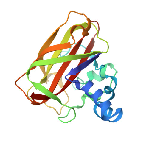
Activity, stability and 3-D structure of the Cu(ii) form of a chitin-active lytic polysaccharide monooxygenase from Bacillus amyloliquefaciens.

Gregory, R.C.Hemsworth, G.R.Turkenburg, J.P.Hart, S.J.Walton, P.H.Davies, G.J.

(2016) Dalton Trans 45: 16904-16912

  • DOI: https://doi.org/10.1039/c6dt02793h
  • Primary Citation of Related Structures:  
    5IJU

  • PubMed Abstract: 

    The enzymatic deconstruction of recalcitrant polysaccharide biomass is central to the conversion of these substrates for societal benefit, such as in biofuels. Traditional models for enzyme-catalysed polysaccharide degradation involved the synergistic action of endo-, exo- and processive glycoside hydrolases working in concert to hydrolyse the substrate. More recently this model has been succeeded by one featuring a newly discovered class of mononuclear copper enzymes: lytic polysaccharide monooxygenases (LPMOs; classified as Auxiliary Activity (AA) enzymes in the CAZy classification). In 2013, the structure of an LPMO from Bacillus amyloliquefaciens, BaAA10, was solved with the Cu centre photoreduced to Cu(i) in the X-ray beam. Here we present the catalytic activity of BaAA10. We show that it is a chitin-active LPMO, active on both α and β chitin, with the Cu(ii) binding with low nM K D , and the substrate greatly increasing the thermal stability of the enzyme. A spiral data collection strategy has been used to facilitate access to the previously unobservable Cu(ii) state of the active centre, revealing a coordination geometry around the copper which is distorted from axial symmetry, consistent with the previous findings from EPR spectroscopy.


  • Organizational Affiliation

    Structural Biology Laboratory, University of York, Heslington, York, YO10 5DD, UK. gideon.davies@york.ac.uk.


Macromolecules
Find similar proteins by:  (by identity cutoff)  |  3D Structure
Entity ID: 1
MoleculeChains Sequence LengthOrganismDetailsImage
BaAA10 Lytic Polysaccharide Monooxygenase
A, B
179Bacillus amyloliquefaciensMutation(s): 0 
Gene Names: RBAM17540BAMF_1859
Entity Groups  
Sequence Clusters30% Identity50% Identity70% Identity90% Identity95% Identity100% Identity
Sequence Annotations
Expand
  • Reference Sequence
Experimental Data & Validation

Experimental Data

  • Method: X-RAY DIFFRACTION
  • Resolution: 1.70 Å
  • R-Value Free: 0.264 
  • R-Value Work: 0.223 
  • R-Value Observed: 0.225 
  • Space Group: P 1 21 1
Unit Cell:
Length ( Å )Angle ( ˚ )
a = 34.779α = 90
b = 73.482β = 100.4
c = 75.457γ = 90
Software Package:
Software NamePurpose
REFMACrefinement
PDB_EXTRACTdata extraction
XDSdata reduction
Aimlessdata scaling
REFMACphasing

Structure Validation

View Full Validation Report


Entry History & Funding Information

Deposition Data


Funding OrganizationLocationGrant Number
Biotechnology and Biological Sciences Research CouncilUnited KingdomBB/I014802/1
Biotechnology and Biological Sciences Research CouncilUnited KingdomBB/L000423
Biotechnology and Biological Sciences Research CouncilUnited KingdomBB/L021633/1

Revision History  (Full details and data files)

  • Version 1.0: 2016-09-21
    Type: Initial release
  • Version 1.1: 2016-10-19
    Changes: Database references
  • Version 1.2: 2016-11-02
    Changes: Database references
  • Version 2.0: 2017-08-30
    Changes: Atomic model, Author supporting evidence
  • Version 2.1: 2024-01-10
    Changes: Data collection, Database references, Derived calculations, Refinement description