5ID2

Asymmetry in the active site of Mycobacterium tuberculosis AhpE upon exposure to Mycothiol


Experimental Data Snapshot

  • Method: X-RAY DIFFRACTION
  • Resolution: 2.43 Å
  • R-Value Free: 0.205 
  • R-Value Work: 0.187 
  • R-Value Observed: 0.188 

wwPDB Validation   3D Report Full Report


This is version 1.3 of the entry. See complete history


Literature

Redox chemistry of Mycobacterium tuberculosis alkylhydroperoxide reductase E (AhpE): Structural and mechanistic insight into a mycoredoxin-1 independent reductive pathway of AhpE via mycothiol

Kumar, A.Balakrishna, A.M.Nartey, W.Manimekalai, M.S.S.Gruber, G.

(2016) Free Radic Biol Med 97: 588-601

  • DOI: https://doi.org/10.1016/j.freeradbiomed.2016.07.007
  • Primary Citation of Related Structures:  
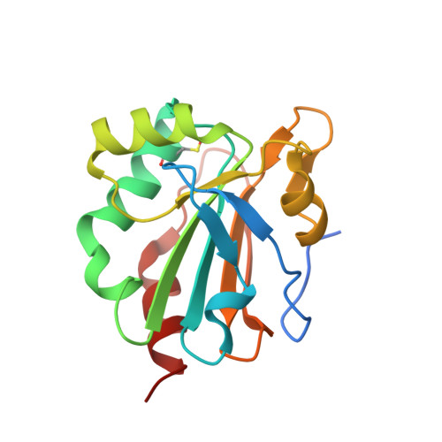
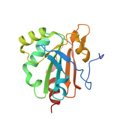
    5ID2

  • PubMed Abstract: 

    Mycobacterium tuberculosis (Mtb) has the ability to persist within the human host for a long time in a dormant stage and re-merges when the immune system is compromised. The pathogenic bacterium employs an elaborate antioxidant defence machinery composed of the mycothiol- and thioredoxin system in addition to a superoxide dismutase, a catalase, and peroxiredoxins (Prxs). Among the family of Peroxiredoxins, Mtb expresses a 1-cysteine peroxiredoxin, known as alkylhydroperoxide reductase E (MtAhpE), and defined as a potential tuberculosis drug target. The reduced MtAhpE (MtAhpE-SH) scavenges peroxides to become converted to MtAhpE-SOH. To provide continuous availability of MtAhpE-SH, MtAhpE-SOH has to become reduced. Here, we used NMR spectroscopy to delineate the reduced (MtAhpE-SH), sulphenic (MtAhpE-SOH) and sulphinic (MtAhpE-SO2H) states of MtAhpE through cysteinyl-labelling, and provide for the first time evidence of a mycothiol-dependent mechanism of MtAhpE reduction. This is confirmed by crystallographic studies, wherein MtAhpE was crystallized in the presence of mycothiol and the structure was solved at 2.43Å resolution. Combined with NMR-studies, the crystallographic structures reveal conformational changes of important residues during the catalytic cycle of MtAhpE. In addition, alterations of the overall protein in solution due to redox modulation are observed by small angle X-ray scattering (SAXS) studies. Finally, by employing SAXS and dynamic light scattering, insight is provided into the most probable physiological oligomeric state of MtAhpE necessary for activity, being also discussed in the context of concerted substrate binding inside the dimeric MtAhpE.


  • Organizational Affiliation

    Nanyang Technological University, School of Biological Sciences, 60 Nanyang Drive, Singapore 637551, Republic of Singapore.


Macromolecules
Find similar proteins by:  (by identity cutoff)  |  3D Structure
Entity ID: 1
MoleculeChains Sequence LengthOrganismDetailsImage
Putative peroxiredoxin Rv2238c
A, D
164Mycobacterium tuberculosis H37RvMutation(s): 0 
Gene Names: Rv2238cMtAhpE
EC: 1.11.1.15
UniProt
Find proteins for P9WIE3 (Mycobacterium tuberculosis (strain ATCC 25618 / H37Rv))
Explore P9WIE3 
Go to UniProtKB:  P9WIE3
Entity Groups  
Sequence Clusters30% Identity50% Identity70% Identity90% Identity95% Identity100% Identity
UniProt GroupP9WIE3
Sequence Annotations
Expand
  • Reference Sequence
Find similar proteins by:  (by identity cutoff)  |  3D Structure
Entity ID: 2
MoleculeChains Sequence LengthOrganismDetailsImage
Putative peroxiredoxin Rv2238c
B, C
164Mycobacterium tuberculosis H37RvMutation(s): 0 
Gene Names: Rv2238cMtAhpE
EC: 1.11.1.15
UniProt
Find proteins for P9WIE3 (Mycobacterium tuberculosis (strain ATCC 25618 / H37Rv))
Explore P9WIE3 
Go to UniProtKB:  P9WIE3
Entity Groups  
Sequence Clusters30% Identity50% Identity70% Identity90% Identity95% Identity100% Identity
UniProt GroupP9WIE3
Sequence Annotations
Expand
  • Reference Sequence
Small Molecules
Modified Residues  1 Unique
IDChains TypeFormula2D DiagramParent
CSO
Query on CSO
A, D
L-PEPTIDE LINKINGC3 H7 N O3 SCYS
Experimental Data & Validation

Experimental Data

  • Method: X-RAY DIFFRACTION
  • Resolution: 2.43 Å
  • R-Value Free: 0.205 
  • R-Value Work: 0.187 
  • R-Value Observed: 0.188 
  • Space Group: P 42
Unit Cell:
Length ( Å )Angle ( ˚ )
a = 146.906α = 90
b = 146.906β = 90
c = 33.616γ = 90
Software Package:
Software NamePurpose
REFMACrefinement
HKL-2000data reduction
HKL-2000data scaling
PHASERphasing

Structure Validation

View Full Validation Report


Entry History 

Deposition Data

Revision History  (Full details and data files)

  • Version 1.0: 2016-08-03
    Type: Initial release
  • Version 1.1: 2016-08-24
    Changes: Database references
  • Version 1.2: 2023-11-08
    Changes: Data collection, Database references, Derived calculations, Refinement description
  • Version 1.3: 2023-11-15
    Changes: Data collection