


Structure of a cysteine hydrolase
Gao, S., Feng, Y.To be published.
Experimental Data Snapshot
wwPDB Validation 3D Report Full Report

Entity ID: 1 | |||||
|---|---|---|---|---|---|
| Molecule | Chains | Sequence Length | Organism | Details | Image |
| Isochorismatase | 187 | Microbacterium hydrocarbonoxydans | Mutation(s): 1 | 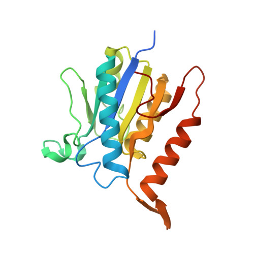 | |
UniProt | |||||
Find proteins for A0A0K0XHU0 (Microbacterium hydrocarbonoxydans) Explore A0A0K0XHU0 Go to UniProtKB: A0A0K0XHU0 | |||||
Entity Groups | |||||
| Sequence Clusters | 30% Identity50% Identity70% Identity90% Identity95% Identity100% Identity | ||||
| UniProt Group | A0A0K0XHU0 | ||||
Sequence AnnotationsExpand | |||||
| |||||
| Ligands 1 Unique | |||||
|---|---|---|---|---|---|
| ID | Chains | Name / Formula / InChI Key | 2D Diagram | 3D Interactions | |
| 66Z Query on 66Z | B [auth A] | (1~{S},4~{R})-3-azabicyclo[2.2.1]hept-5-en-2-one C6 H7 N O DDUFYKNOXPZZIW-UHNVWZDZSA-N |  | ||
| Length ( Å ) | Angle ( ˚ ) |
|---|---|
| a = 69.427 | α = 90 |
| b = 69.427 | β = 90 |
| c = 87.159 | γ = 120 |
| Software Name | Purpose |
|---|---|
| PHENIX | refinement |
| HKL-2000 | data reduction |
| SCALEPACK | data scaling |
| PHASER | phasing |
RCSB PDB (citation) is hosted by
RCSB PDB is a member of the

