5HJO

Murine endoplasmic reticulum alpha-glucosidase II with bound substrate analogue


Experimental Data Snapshot

  • Method: X-RAY DIFFRACTION
  • Resolution: 2.29 Å
  • R-Value Free: 0.236 
  • R-Value Work: 0.204 
  • R-Value Observed: 0.206 

wwPDB Validation   3D Report Full Report


This is version 2.0 of the entry. See complete history


Literature

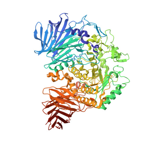
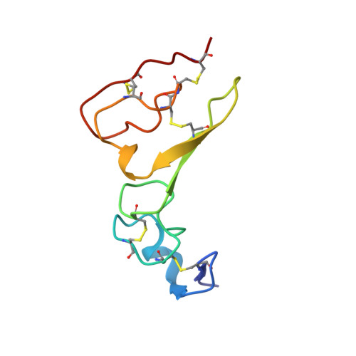
Structures of mammalian ER alpha-glucosidase II capture the binding modes of broad-spectrum iminosugar antivirals.

Caputo, A.T.Alonzi, D.S.Marti, L.Reca, I.B.Kiappes, J.L.Struwe, W.B.Cross, A.Basu, S.Lowe, E.D.Darlot, B.Santino, A.Roversi, P.Zitzmann, N.

(2016) Proc Natl Acad Sci U S A 113: E4630-E4638

  • DOI: https://doi.org/10.1073/pnas.1604463113
  • Primary Citation of Related Structures:  
    5F0E, 5H9O, 5HJO, 5HJR, 5IED, 5IEE, 5IEF, 5IEG

  • PubMed Abstract: 

    The biosynthesis of enveloped viruses depends heavily on the host cell endoplasmic reticulum (ER) glycoprotein quality control (QC) machinery. This dependency exceeds the dependency of host glycoproteins, offering a window for the targeting of ERQC for the development of broad-spectrum antivirals. We determined small-angle X-ray scattering (SAXS) and crystal structures of the main ERQC enzyme, ER α-glucosidase II (α-GluII; from mouse), alone and in complex with key ligands of its catalytic cycle and antiviral iminosugars, including two that are in clinical trials for the treatment of dengue fever. The SAXS data capture the enzyme's quaternary structure and suggest a conformational rearrangement is needed for the simultaneous binding of a monoglucosylated glycan to both subunits. The X-ray structures with key catalytic cycle intermediates highlight that an insertion between the +1 and +2 subsites contributes to the enzyme's activity and substrate specificity, and reveal that the presence of d-mannose at the +1 subsite renders the acid catalyst less efficient during the cleavage of the monoglucosylated substrate. The complexes with iminosugar antivirals suggest that inhibitors targeting a conserved ring of aromatic residues between the α-GluII +1 and +2 subsites would have increased potency and selectivity, thus providing a template for further rational drug design.


  • Organizational Affiliation

    Oxford Glycobiology Institute, Department of Biochemistry, University of Oxford, Oxford OX1 3QU, United Kingdom;


Macromolecules
Find similar proteins by:  (by identity cutoff)  |  3D Structure
Entity ID: 1
MoleculeChains Sequence LengthOrganismDetailsImage
Neutral alpha-glucosidase AB
A, C
857Mus musculusMutation(s): 0 
Gene Names: GanabG2anKiaa0088
EC: 3.2.1.84
UniProt
Find proteins for Q8BHN3 (Mus musculus)
Explore Q8BHN3 
Go to UniProtKB:  Q8BHN3
Entity Groups  
Sequence Clusters30% Identity50% Identity70% Identity90% Identity95% Identity100% Identity
UniProt GroupQ8BHN3
Sequence Annotations
Expand
  • Reference Sequence
Find similar proteins by:  (by identity cutoff)  |  3D Structure
Entity ID: 2
MoleculeChains Sequence LengthOrganismDetailsImage
Glucosidase 2 subunit beta
B, D
83Mus musculusMutation(s): 0 
Gene Names: Prkcsh
UniProt & NIH Common Fund Data Resources
Find proteins for O08795 (Mus musculus)
Explore O08795 
Go to UniProtKB:  O08795
IMPC:  MGI:107877
Entity Groups  
Sequence Clusters30% Identity50% Identity70% Identity90% Identity95% Identity100% Identity
UniProt GroupO08795
Sequence Annotations
Expand
  • Reference Sequence
Oligosaccharides

Help

Entity ID: 3
MoleculeChains Length2D Diagram Glycosylation3D Interactions
alpha-D-mannopyranose-(1-3)-beta-D-mannopyranose-(1-4)-2-acetamido-2-deoxy-beta-D-glucopyranose-(1-4)-2-acetamido-2-deoxy-beta-D-glucopyranose
E, F
4N-Glycosylation
Glycosylation Resources
GlyTouCan:  G81315DD
GlyCosmos:  G81315DD
GlyGen:  G81315DD
Entity ID: 4
MoleculeChains Length2D Diagram Glycosylation3D Interactions
2-deoxy-alpha-D-arabino-hexopyranose-(1-3)-D-glucal
G, H
2N/A
Glycosylation Resources
GlyTouCan:  G58893KD
GlyCosmos:  G58893KD
Small Molecules
Ligands 5 Unique
IDChains Name / Formula / InChI Key2D Diagram3D Interactions
DGO
Query on DGO

Download Ideal Coordinates CCD File 
JA [auth D],
U [auth B]
D-glucal
C6 H10 O4
YVECGMZCTULTIS-PBXRRBTRSA-N
EDO
Query on EDO

Download Ideal Coordinates CCD File 
J [auth A],
Y [auth C]
1,2-ETHANEDIOL
C2 H6 O2
LYCAIKOWRPUZTN-UHFFFAOYSA-N
ACT
Query on ACT

Download Ideal Coordinates CCD File 
I [auth A],
X [auth C]
ACETATE ION
C2 H3 O2
QTBSBXVTEAMEQO-UHFFFAOYSA-M
FMT
Query on FMT

Download Ideal Coordinates CCD File 
AA [auth C]
BA [auth C]
CA [auth C]
DA [auth C]
EA [auth C]
AA [auth C],
BA [auth C],
CA [auth C],
DA [auth C],
EA [auth C],
FA [auth C],
GA [auth C],
HA [auth C],
IA [auth C],
K [auth A],
L [auth A],
M [auth A],
N [auth A],
O [auth A],
P [auth A],
Q [auth A],
R [auth A],
S [auth A],
T [auth A],
Z [auth C]
FORMIC ACID
C H2 O2
BDAGIHXWWSANSR-UHFFFAOYSA-N
CA
Query on CA

Download Ideal Coordinates CCD File 
KA [auth D],
LA [auth D],
V [auth B],
W [auth B]
CALCIUM ION
Ca
BHPQYMZQTOCNFJ-UHFFFAOYSA-N
Experimental Data & Validation

Experimental Data

  • Method: X-RAY DIFFRACTION
  • Resolution: 2.29 Å
  • R-Value Free: 0.236 
  • R-Value Work: 0.204 
  • R-Value Observed: 0.206 
  • Space Group: P 21 21 21
Unit Cell:
Length ( Å )Angle ( ˚ )
a = 127.05α = 90
b = 176.44β = 90
c = 103.48γ = 90
Software Package:
Software NamePurpose
BUSTERrefinement
autoPROCdata reduction
Aimlessdata scaling
PHASERphasing

Structure Validation

View Full Validation Report


Entry History 

Deposition Data

Revision History  (Full details and data files)

  • Version 1.0: 2016-07-27
    Type: Initial release
  • Version 1.1: 2016-08-03
    Changes: Database references
  • Version 2.0: 2020-07-29
    Type: Remediation
    Reason: Carbohydrate remediation
    Changes: Atomic model, Data collection, Derived calculations, Non-polymer description, Refinement description, Source and taxonomy, Structure summary