Substrate Binding Mode and Molecular Basis of a Specificity Switch in Oxalate Decarboxylase.
Zhu, W., Easthon, L.M., Reinhardt, L.A., Tu, C., Cohen, S.E., Silverman, D.N., Allen, K.N., Richards, N.G.(2016) Biochemistry 55: 2163-2173
- PubMed: 27014926
- DOI: https://doi.org/10.1021/acs.biochem.6b00043
- Primary Citation of Related Structures:
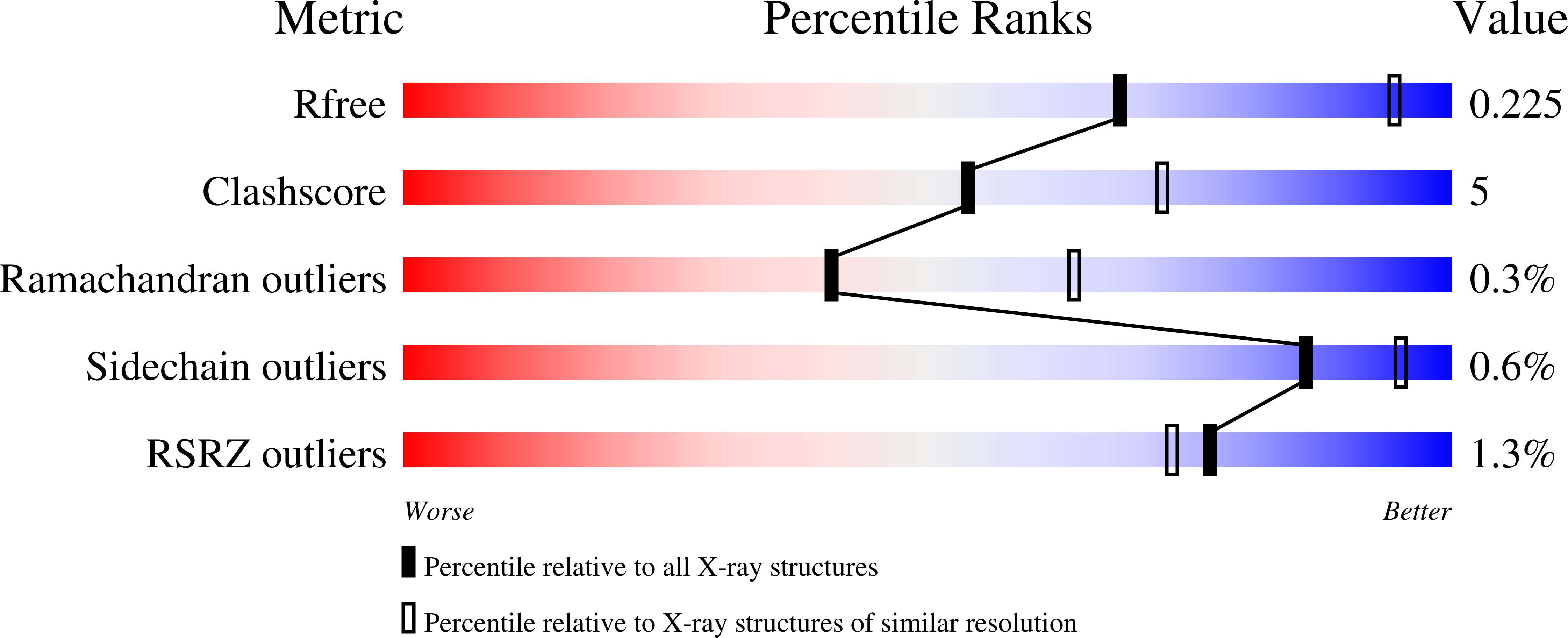
5HI0 - PubMed Abstract:
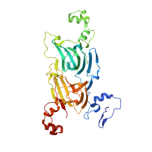
Oxalate decarboxylase (OxDC) catalyzes the conversion of oxalate into formate and carbon dioxide in a remarkable reaction that requires manganese and dioxygen. Previous studies have shown that replacing an active-site loop segment Ser(161)-Glu(162)-Asn(163)-Ser(164) in the N-terminal domain of OxDC with the cognate residues Asp(161)-Ala(162)-Ser-(163)-Asn(164) of an evolutionarily related, Mn-dependent oxalate oxidase gives a chimeric variant (DASN) that exhibits significantly increased oxidase activity. The mechanistic basis for this change in activity has now been investigated using membrane inlet mass spectrometry (MIMS) and isotope effect (IE) measurements. Quantitative analysis of the reaction stoichiometry as a function of oxalate concentration, as determined by MIMS, suggests that the increased oxidase activity of the DASN OxDC variant is associated with only a small fraction of the enzyme molecules in solution. In addition, IE measurements show that C-C bond cleavage in the DASN OxDC variant proceeds via the same mechanism as in the wild-type enzyme, even though the Glu(162) side chain is absent. Thus, replacement of the loop residues does not modulate the chemistry of the enzyme-bound Mn(II) ion. Taken together, these results raise the possibility that the observed oxidase activity of the DASN OxDC variant arises from an increased level of access of the solvent to the active site during catalysis, implying that the functional role of Glu(162) is to control loop conformation. A 2.6 Å resolution X-ray crystal structure of a complex between oxalate and the Co(II)-substituted ΔE162 OxDC variant, in which Glu(162) has been deleted from the active site loop, reveals the likely mode by which the substrate coordinates the catalytically active Mn ion prior to C-C bond cleavage. The "end-on" conformation of oxalate observed in the structure is consistent with the previously published V/K IE data and provides an empty coordination site for the dioxygen ligand that is thought to mediate the formation of Mn(III) for catalysis upon substrate binding.
Organizational Affiliation:
Department of Chemistry & Chemical Biology, Indiana University-Purdue University Indianapolis , Indianapolis, Indiana 46202, United States.