5HCH

X-ray structure of a lectin-bound DNA duplex containing an unnatural phenanthrenyl pair


Experimental Data Snapshot

  • Method: X-RAY DIFFRACTION
  • Resolution: 2.90 Å
  • R-Value Free: 0.257 
  • R-Value Work: 0.227 
  • R-Value Observed: 0.230 

wwPDB Validation   3D Report Full Report


Ligand Structure Quality Assessment 


This is version 2.1 of the entry. See complete history


Literature

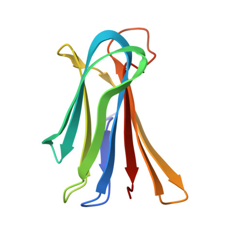
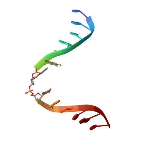
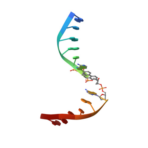
X-ray structure of a lectin-bound DNA duplex containing an unnatural phenanthrenyl pair.

Roethlisberger, P.Istrate, A.Marcaida Lopez, M.J.Visini, R.Stocker, A.Reymond, J.L.Leumann, C.J.

(2016) Chem Commun (Camb) 52: 4749-4752

  • DOI: https://doi.org/10.1039/c6cc00374e
  • Primary Citation of Related Structures:  
    5HCH

  • PubMed Abstract: 

    DNA duplexes containing unnatural base-pair surrogates are attractive biomolecular nanomaterials with potentially beneficial photophysical or electronic properties. Herein we report the first X-ray structure of a duplex containing a phen-pair in the center of the double helix in a zipper like stacking arrangement.


  • Organizational Affiliation

    Department of Chemistry and Biochemistry, University of Bern, Freiestrasse 3, 3012 Bern, Switzerland. christian.leumann@dcb.unibe.ch.


Macromolecules

Find similar proteins by:  (by identity cutoff)  |  3D Structure
Entity ID: 1
MoleculeChains Sequence LengthOrganismDetailsImage
Fucose-binding lectin114Pseudomonas aeruginosaMutation(s): 0 
Gene Names: 
UniProt
Find proteins for Q9HYN5 (Pseudomonas aeruginosa (strain ATCC 15692 / DSM 22644 / CIP 104116 / JCM 14847 / LMG 12228 / 1C / PRS 101 / PAO1))
Explore Q9HYN5 
Go to UniProtKB:  Q9HYN5
Entity Groups  
Sequence Clusters30% Identity50% Identity70% Identity90% Identity95% Identity100% Identity
UniProt GroupQ9HYN5
Sequence Annotations
Expand
  • Reference Sequence

Find similar nucleic acids by:  Sequence   |   3D Structure  

Entity ID: 2
MoleculeChains LengthOrganismImage
DNA (5'-D(*CP*GP*CP*AP*TP*TP*(DF)P*TP*AP*TP*CP*GP*C)-3')13synthetic construct
Sequence Annotations
Expand
  • Reference Sequence

Find similar nucleic acids by:  Sequence   |   3D Structure  

Entity ID: 3
MoleculeChains LengthOrganismImage
DNA (5'-D(P*GP*CP*GP*AP*TP*AP*(DF)P*AP*AP*TP*GP*CP*G)-3')C [auth E]13synthetic construct
Sequence Annotations
Expand
  • Reference Sequence
Experimental Data & Validation

Experimental Data

  • Method: X-RAY DIFFRACTION
  • Resolution: 2.90 Å
  • R-Value Free: 0.257 
  • R-Value Work: 0.227 
  • R-Value Observed: 0.230 
  • Space Group: P 62 2 2
Unit Cell:
Length ( Å )Angle ( ˚ )
a = 60.451α = 90
b = 60.451β = 90
c = 204.828γ = 120
Software Package:
Software NamePurpose
PHENIXrefinement
PHASERphasing
PDB_EXTRACTdata extraction
XDSdata reduction
XSCALEdata scaling

Structure Validation

View Full Validation Report


Ligand Structure Quality Assessment 


Entry History & Funding Information

Deposition Data


Funding OrganizationLocationGrant Number
Swiss National Science FoundationSwitzerland20020_146646

Revision History  (Full details and data files)

  • Version 1.0: 2016-03-02
    Type: Initial release
  • Version 1.1: 2016-03-30
    Changes: Database references
  • Version 1.2: 2017-09-13
    Changes: Author supporting evidence, Derived calculations
  • Version 2.0: 2020-07-29
    Type: Remediation
    Reason: Carbohydrate remediation
    Changes: Atomic model, Data collection, Derived calculations
  • Version 2.1: 2024-01-10
    Changes: Data collection, Database references, Refinement description