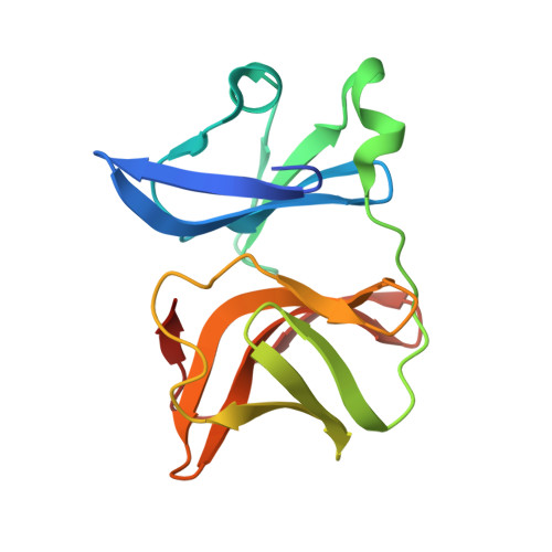
5H23

Crystal structure of Chikungunya virus capsid protein


Experimental Data Snapshot

  • Method: X-RAY DIFFRACTION
  • Resolution: 2.20 Å
  • R-Value Free: 0.261 
  • R-Value Work: 0.176 
  • R-Value Observed: 0.180 

wwPDB Validation   3D Report Full Report


Ligand Structure Quality Assessment 


This is version 1.3 of the entry. See complete history


Literature

Structure-function insights into chikungunya virus capsid protein: Small molecules targeting capsid hydrophobic pocket.

Sharma, R.Kesari, P.Kumar, P.Tomar, S.

(2018) Virology 515: 223-234

  • DOI: https://doi.org/10.1016/j.virol.2017.12.020
  • Primary Citation of Related Structures:  
    5H23

  • PubMed Abstract: 

    The crystal structure of chikungunya (CHIKV) virus capsid protease domain has been determined at 2.2Å. Structure reveals a chymotrypsin-like protease fold with a conserved hydrophobic pocket in CHIKV capsid protein (CP) for interaction with the cytoplasmic tail of E2 (cdE2) similar to the capsid protein of other alphaviruses. Molecular contacts between CP-cdE2 were determined by fitting structures of CHIKV CP and cdE2 into the cryo-EM map of Venezuelan equine encephalitis virus (VEEV). Binding of (S)-(+)-Mandelic acid (MDA) and Ethyl 3-aminobenzoate (EAB) to the hydrophobic pocket of CP was evaluated by molecular docking. Surface plasmon resonance (SPR) and fluorescence spectroscopy experiments confirmed MDA and EAB binding to the CP. The binding constants (K D ) obtained from SPR for MDA and EAB were 1.2 × 10 -3 M and 0.2 × 10 -9 M, respectively. This study adds to the understanding of chikungunya virus structural proteins and may serve as the basis for antiviral development against chikungunya disease.


  • Organizational Affiliation

    Department of Biotechnology, Indian Institute of Technology Roorkee, Roorkee 247667, India.


Macromolecules
Find similar proteins by:  (by identity cutoff)  |  3D Structure
Entity ID: 1
MoleculeChains Sequence LengthOrganismDetailsImage
Capsid Protein
A, B
156Chikungunya virusMutation(s): 0 
UniProt
Find proteins for P0DOK1 (Chikungunya virus (strain S27-African prototype))
Explore P0DOK1 
Go to UniProtKB:  P0DOK1
Entity Groups  
Sequence Clusters30% Identity50% Identity70% Identity90% Identity95% Identity100% Identity
UniProt GroupP0DOK1
Sequence Annotations
Expand
  • Reference Sequence
Small Molecules
Ligands 4 Unique
IDChains Name / Formula / InChI Key2D Diagram3D Interactions
BOG
Query on BOG

Download Ideal Coordinates CCD File 
F [auth A],
K [auth B]
octyl beta-D-glucopyranoside
C14 H28 O6
HEGSGKPQLMEBJL-RKQHYHRCSA-N
PEG
Query on PEG

Download Ideal Coordinates CCD File 
D [auth A]DI(HYDROXYETHYL)ETHER
C4 H10 O3
MTHSVFCYNBDYFN-UHFFFAOYSA-N
GOL
Query on GOL

Download Ideal Coordinates CCD File 
E [auth A],
J [auth B]
GLYCEROL
C3 H8 O3
PEDCQBHIVMGVHV-UHFFFAOYSA-N
EDO
Query on EDO

Download Ideal Coordinates CCD File 
C [auth A],
G [auth B],
H [auth B],
I [auth B]
1,2-ETHANEDIOL
C2 H6 O2
LYCAIKOWRPUZTN-UHFFFAOYSA-N
Experimental Data & Validation

Experimental Data

  • Method: X-RAY DIFFRACTION
  • Resolution: 2.20 Å
  • R-Value Free: 0.261 
  • R-Value Work: 0.176 
  • R-Value Observed: 0.180 
  • Space Group: P 1
Unit Cell:
Length ( Å )Angle ( ˚ )
a = 33.307α = 92.36
b = 39.096β = 91.18
c = 59.261γ = 103
Software Package:
Software NamePurpose
PHENIXrefinement
DENZOdata reduction
SCALEPACKdata scaling
MOLREPphasing

Structure Validation

View Full Validation Report


Ligand Structure Quality Assessment 


Entry History 

Deposition Data

Revision History  (Full details and data files)

  • Version 1.0: 2018-03-14
    Type: Initial release
  • Version 1.1: 2019-04-03
    Changes: Data collection, Database references
  • Version 1.2: 2020-07-29
    Type: Remediation
    Reason: Carbohydrate remediation
    Changes: Data collection, Derived calculations, Structure summary
  • Version 1.3: 2023-11-08
    Changes: Data collection, Database references, Refinement description, Structure summary