5GQO

Structure of the second Single Stranded DNA Binding protein (SSBb) from Mycobacterium smegmatis


Experimental Data Snapshot

  • Method: X-RAY DIFFRACTION
  • Resolution: 2.50 Å
  • R-Value Free: 0.256 
  • R-Value Work: 0.211 
  • R-Value Observed: 0.213 

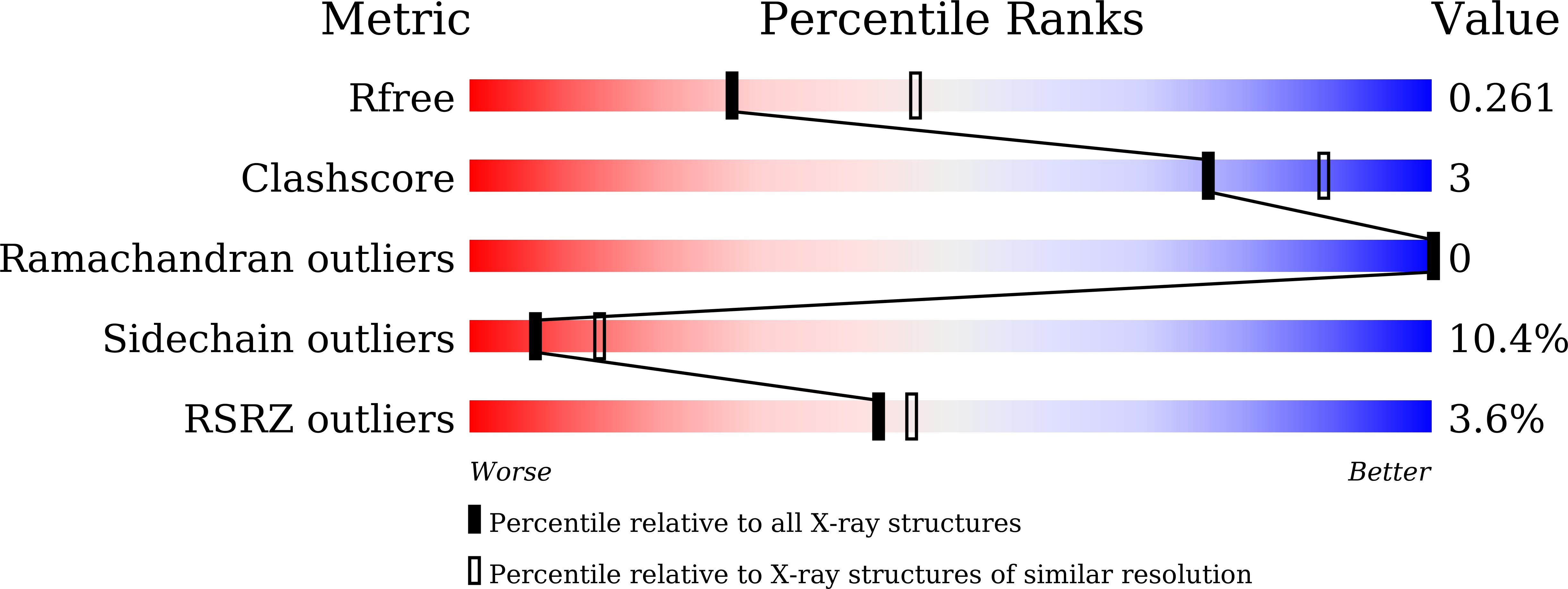
wwPDB Validation   3D Report Full Report


This is version 1.2 of the entry. See complete history


Literature

Structure of the second Single Stranded DNA Binding protein (SSBb) from Mycobacterium smegmatis.

Singh, A.Varshney, U.Vijayan, M.

(2016) J Struct Biol 196: 448-454

  • DOI: https://doi.org/10.1016/j.jsb.2016.09.012
  • Primary Citation of Related Structures:  
    5GQO

  • PubMed Abstract: 

    All mycobacteria with sequenced genomes, except M. leprae, have a second Single Stranded DNA Binding protein (SSBb) in addition to the canonical one (SSBa). This paralogue from M. smegmatis (MsSSBb) has been cloned, expressed and purified. The protein, which is probably involved in stress response, has been crystallized and X-ray analyzed in the first structure elucidation of a mycobacterial SSBb. In spite of the low sequence identity between SSBas and SSBbs in mycobacteria, the tertiary and quaternary structure of the DNA binding domain of MsSSBb is similar to that observed in mycobacterial SSBas. In particular, the quaternary structure is 'clamped' using a C-terminal stretch of the N-domain, which endows the tetrameric molecule with additional stability and its characteristic shape. Comparison involving available, rather limited, structural data on SSBbs from other sources, appears to suggest that SSBbs could exhibit higher structural variability than SSBas do.


  • Organizational Affiliation

    Molecular Biophysics Unit, Indian Institute of Science, Bangalore 560012, Karnataka, India.


Macromolecules
Find similar proteins by:  (by identity cutoff)  |  3D Structure
Entity ID: 1
MoleculeChains Sequence LengthOrganismDetailsImage
Single-stranded DNA-binding protein
A, B
190Mycolicibacterium smegmatis MC2 155Mutation(s): 0 
Gene Names: MSMEG_4701
UniProt
Find proteins for A0R1C4 (Mycolicibacterium smegmatis (strain ATCC 700084 / mc(2)155))
Explore A0R1C4 
Go to UniProtKB:  A0R1C4
Entity Groups  
Sequence Clusters30% Identity50% Identity70% Identity90% Identity95% Identity100% Identity
UniProt GroupA0R1C4
Sequence Annotations
Expand
  • Reference Sequence
Small Molecules
Ligands 1 Unique
IDChains Name / Formula / InChI Key2D Diagram3D Interactions
CA
Query on CA

Download Ideal Coordinates CCD File 
C [auth A]CALCIUM ION
Ca
BHPQYMZQTOCNFJ-UHFFFAOYSA-N
Experimental Data & Validation

Experimental Data

  • Method: X-RAY DIFFRACTION
  • Resolution: 2.50 Å
  • R-Value Free: 0.256 
  • R-Value Work: 0.211 
  • R-Value Observed: 0.213 
  • Space Group: P 65 2 2
Unit Cell:
Length ( Å )Angle ( ˚ )
a = 73.61α = 90
b = 73.61β = 90
c = 216.21γ = 120
Software Package:
Software NamePurpose
REFMACrefinement
iMOSFLMdata reduction
SCALAdata scaling
PHASERphasing

Structure Validation

View Full Validation Report


Entry History 

Deposition Data

Revision History  (Full details and data files)

  • Version 1.0: 2016-10-12
    Type: Initial release
  • Version 1.1: 2017-12-06
    Changes: Database references, Derived calculations
  • Version 1.2: 2023-11-08
    Changes: Data collection, Database references, Derived calculations, Refinement description