5G2E

Structure of the Nap1 H2A H2B complex


Experimental Data Snapshot

  • Method: X-RAY DIFFRACTION
  • Resolution: 6.70 Å
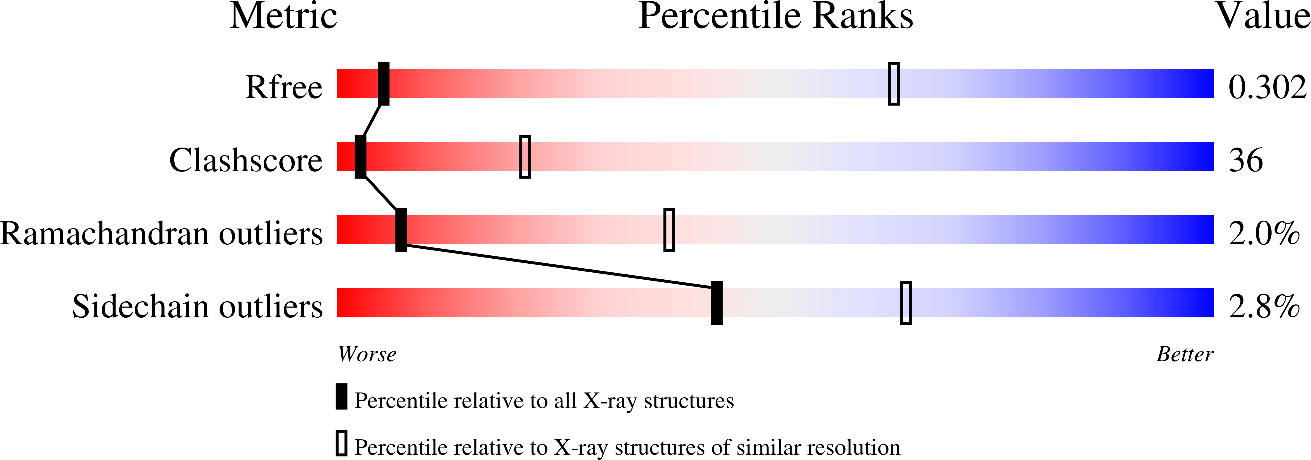
  • R-Value Free: 0.310 
  • R-Value Work: 0.263 
  • R-Value Observed: 0.263 

wwPDB Validation   3D Report Full Report


This is version 1.1 of the entry. See complete history


Literature

Structural Evidence for Nap1-Dependent H2A-H2B Deposition and Nucleosome Assembly.

Aguilar-Gurrieri, C.Larabi, A.Vinayachandran, V.Patel, N.A.Yen, K.Reja, R.Ebong, I.Schoehn, G.Robinson, C.V.Pugh, B.F.Panne, D.

(2016) EMBO J 35: 1465

  • DOI: https://doi.org/10.15252/embj.201694105
  • Primary Citation of Related Structures:  
    5G2E

  • PubMed Abstract: 

    Nap1 is a histone chaperone involved in the nuclear import of H2A-H2B and nucleosome assembly. Here, we report the crystal structure of Nap1 bound to H2A-H2B together with in vitro and in vivo functional studies that elucidate the principles underlying Nap1-mediated H2A-H2B chaperoning and nucleosome assembly. A Nap1 dimer provides an acidic binding surface and asymmetrically engages a single H2A-H2B heterodimer. Oligomerization of the Nap1-H2A-H2B complex results in burial of surfaces required for deposition of H2A-H2B into nucleosomes. Chromatin immunoprecipitation-exonuclease (ChIP-exo) analysis shows that Nap1 is required for H2A-H2B deposition across the genome. Mutants that interfere with Nap1 oligomerization exhibit severe nucleosome assembly defects showing that oligomerization is essential for the chaperone function. These findings establish the molecular basis for Nap1-mediated H2A-H2B deposition and nucleosome assembly.


  • Organizational Affiliation

    European Molecular Biology Laboratory, Grenoble, France Unit for Virus Host-Cell Interactions, Univ. Grenoble Alpes-EMBL-CNRS, Grenoble, France.


Macromolecules
Find similar proteins by:  (by identity cutoff)  |  3D Structure
Entity ID: 1
MoleculeChains Sequence LengthOrganismDetailsImage
NUCLEOSOME ASSEMBLY PROTEIN
A, B, E, F, I
A, B, E, F, I, J, M, N, Q, R, U, V
310Saccharomyces cerevisiaeMutation(s): 0 
UniProt
Find proteins for P25293 (Saccharomyces cerevisiae (strain ATCC 204508 / S288c))
Explore P25293 
Go to UniProtKB:  P25293
Entity Groups  
Sequence Clusters30% Identity50% Identity70% Identity90% Identity95% Identity100% Identity
UniProt GroupP25293
Sequence Annotations
Expand
  • Reference Sequence
Find similar proteins by:  (by identity cutoff)  |  3D Structure
Entity ID: 2
MoleculeChains Sequence LengthOrganismDetailsImage
HISTONE H2A TYPE 1
C, G, K, O, S
C, G, K, O, S, W
107Xenopus laevisMutation(s): 0 
UniProt
Find proteins for P06897 (Xenopus laevis)
Explore P06897 
Go to UniProtKB:  P06897
Entity Groups  
Sequence Clusters30% Identity50% Identity70% Identity90% Identity95% Identity100% Identity
UniProt GroupP06897
Sequence Annotations
Expand
  • Reference Sequence
Find similar proteins by:  (by identity cutoff)  |  3D Structure
Entity ID: 3
MoleculeChains Sequence LengthOrganismDetailsImage
HISTONE H2B 1.1
D, H, L, P, T
D, H, L, P, T, X
100Xenopus laevisMutation(s): 0 
UniProt
Find proteins for P02281 (Xenopus laevis)
Explore P02281 
Go to UniProtKB:  P02281
Entity Groups  
Sequence Clusters30% Identity50% Identity70% Identity90% Identity95% Identity100% Identity
UniProt GroupP02281
Sequence Annotations
Expand
  • Reference Sequence
Experimental Data & Validation

Experimental Data

  • Method: X-RAY DIFFRACTION
  • Resolution: 6.70 Å
  • R-Value Free: 0.310 
  • R-Value Work: 0.263 
  • R-Value Observed: 0.263 
  • Space Group: P 1 21 1
Unit Cell:
Length ( Å )Angle ( ˚ )
a = 111.47α = 90
b = 211.13β = 99.72
c = 126.57γ = 90
Software Package:
Software NamePurpose
CNSrefinement
XDSdata reduction
XDSdata scaling
PHASERphasing

Structure Validation

View Full Validation Report


Entry History 

Deposition Data

Revision History  (Full details and data files)

  • Version 1.0: 2016-08-03
    Type: Initial release
  • Version 1.1: 2024-01-10
    Changes: Data collection, Database references, Other, Refinement description