5G09

The crystal structure of a S-selective transaminase from Bacillus megaterium bound with R-alpha-methylbenzylamine


Experimental Data Snapshot

  • Method: X-RAY DIFFRACTION
  • Resolution: 1.90 Å
  • R-Value Free: 0.203 
  • R-Value Work: 0.166 
  • R-Value Observed: 0.167 

wwPDB Validation   3D Report Full Report


Ligand Structure Quality Assessment 


This is version 1.2 of the entry. See complete history


Literature

Structural Basis of Substrate Range and Enantioselectivity of Two S-Selective Omega- Transaminases

Van Oosterwijk, N.Willies, S.Hekelaar, J.Terwisscha Van Scheltinga, A.C.Turner, N.J.Dijkstra, B.W.

(2016) Biochemistry 55: 4422

  • DOI: https://doi.org/10.1021/acs.biochem.6b00370
  • Primary Citation of Related Structures:  
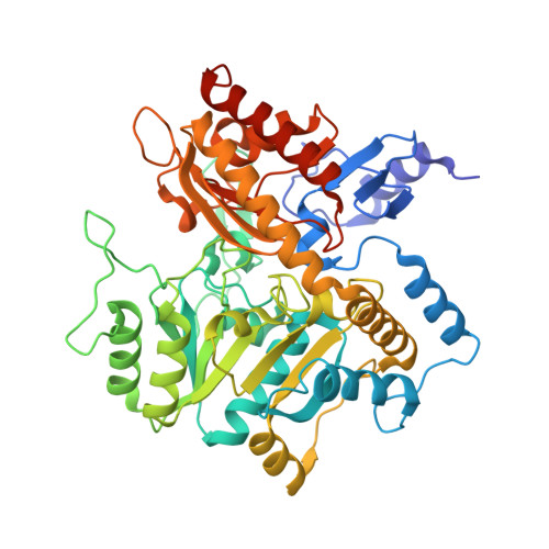
    5G09, 5G0A, 5G2P, 5G2Q

  • PubMed Abstract: 

    ω-Transaminases are enzymes that can introduce an amino group in industrially interesting compounds. We determined crystal structures of two (S)-selective ω-transaminases, one from Arthrobacter sp. (Ars-ωTA) and one from Bacillus megaterium (BM-ωTA), which have 95% identical sequences but somewhat different activity profiles. Substrate profiling measurements using a range of (R)- and (S)-substrates showed that both enzymes have a preference for substrates with large, flat cyclic side groups, for which the activity of BM-ωTA is generally somewhat higher. BM-ωTA has a preference for (S)-3,3-dimethyl-2-butylamine significantly stronger than that of Ars-ωTA, as well as a weaker enantiopreference for 1-cyclopropylethylamine. The crystal structures showed that, as expected for (S)-selective transaminases, both enzymes have the typical transaminase type I fold and have spacious active sites to accommodate largish substrates. A structure of BM-ωTA with bound (R)-α-methylbenzylamine explains the enzymes' preference for (S)-substrates. Site-directed mutagenesis experiments revealed that the presence of a tyrosine, instead of a cysteine, at position 60 increases the relative activities on several small substrates. A structure of Ars-ωTA with bound l-Ala revealed that the Arg442 side chain has been repositioned to bind the l-Ala carboxylate. Compared to the arginine switch residue in other transaminases, Arg442 is shifted by six residues in the amino acid sequence, which appears to be a consequence of extra loops near the active site that narrow the entrance to the active site.


  • Organizational Affiliation

    Laboratory of Biophysical Chemistry, Groningen Biomolecular Sciences and Biotechnology Institute (GBB), University of Groningen , Nijenborgh 7, 9747 AG Groningen, The Netherlands.


Macromolecules
Find similar proteins by:  (by identity cutoff)  |  3D Structure
Entity ID: 1
MoleculeChains Sequence LengthOrganismDetailsImage
TRANSAMINASE
A, B, C, D
483Priestia megateriumMutation(s): 0 
EC: 2.6.1
UniProt
Find proteins for A0A1C7D190 (Priestia megaterium)
Explore A0A1C7D190 
Go to UniProtKB:  A0A1C7D190
Entity Groups  
Sequence Clusters30% Identity50% Identity70% Identity90% Identity95% Identity100% Identity
UniProt GroupA0A1C7D190
Sequence Annotations
Expand
  • Reference Sequence
Small Molecules
Ligands 5 Unique
IDChains Name / Formula / InChI Key2D Diagram3D Interactions
6DF
Query on 6DF

Download Ideal Coordinates CCD File 
E [auth A],
J [auth B],
M [auth C],
R [auth D]
[6-methyl-5-oxidanyl-4-[(~{E})-[(1~{R})-1-phenylethyl]iminomethyl]pyridin-3-yl]methyl dihydrogen phosphate
C16 H19 N2 O5 P
ZBWJIKYNMPWLJE-PBFYJAPKSA-N
1PE
Query on 1PE

Download Ideal Coordinates CCD File 
H [auth A]PENTAETHYLENE GLYCOL
C10 H22 O6
JLFNLZLINWHATN-UHFFFAOYSA-N
PG4
Query on PG4

Download Ideal Coordinates CCD File 
Q [auth D],
T [auth D]
TETRAETHYLENE GLYCOL
C8 H18 O5
UWHCKJMYHZGTIT-UHFFFAOYSA-N
PGE
Query on PGE

Download Ideal Coordinates CCD File 
F [auth A]
G [auth A]
I [auth A]
K [auth B]
L [auth B]
F [auth A],
G [auth A],
I [auth A],
K [auth B],
L [auth B],
N [auth C],
O [auth C],
P [auth C]
TRIETHYLENE GLYCOL
C6 H14 O4
ZIBGPFATKBEMQZ-UHFFFAOYSA-N
PEG
Query on PEG

Download Ideal Coordinates CCD File 
S [auth D]DI(HYDROXYETHYL)ETHER
C4 H10 O3
MTHSVFCYNBDYFN-UHFFFAOYSA-N
Experimental Data & Validation

Experimental Data

  • Method: X-RAY DIFFRACTION
  • Resolution: 1.90 Å
  • R-Value Free: 0.203 
  • R-Value Work: 0.166 
  • R-Value Observed: 0.167 
  • Space Group: P 21 21 21
Unit Cell:
Length ( Å )Angle ( ˚ )
a = 119.317α = 90
b = 124.593β = 90
c = 126.972γ = 90
Software Package:
Software NamePurpose
REFMACrefinement
XDSdata reduction
XDSdata scaling
PHASERphasing

Structure Validation

View Full Validation Report


Ligand Structure Quality Assessment 


Entry History 

Deposition Data

Revision History  (Full details and data files)

  • Version 1.0: 2016-07-27
    Type: Initial release
  • Version 1.1: 2016-08-24
    Changes: Database references
  • Version 1.2: 2024-01-10
    Changes: Data collection, Database references, Derived calculations, Other, Refinement description