5FV0

The cytoplasmic domain of EssC


Experimental Data Snapshot

  • Method: X-RAY DIFFRACTION
  • Resolution: 2.91 Å
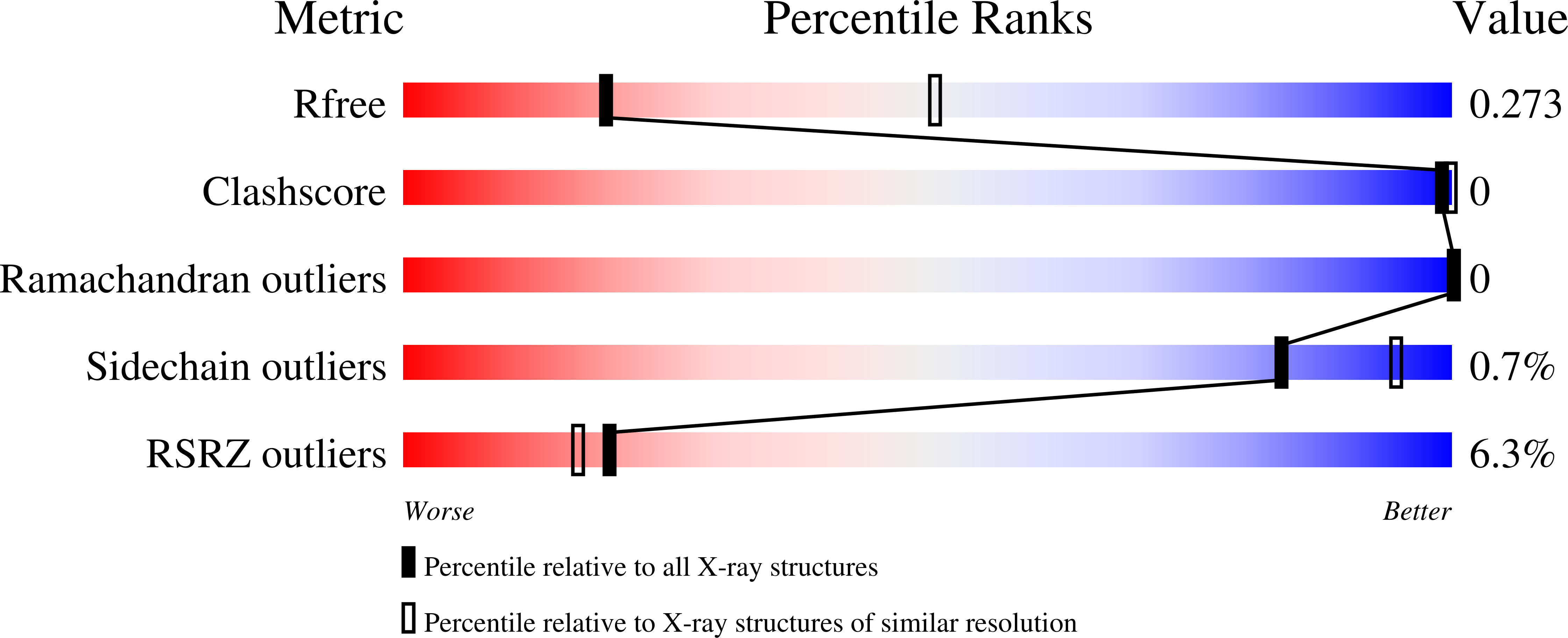
  • R-Value Free: 0.275 
  • R-Value Work: 0.261 
  • R-Value Observed: 0.262 

wwPDB Validation   3D Report Full Report


Ligand Structure Quality Assessment 


This is version 1.2 of the entry. See complete history


Literature

EssC: domain structures inform on the elusive translocation channel in the Type VII secretion system.

Zoltner, M.Ng, W.M.Money, J.J.Fyfe, P.K.Kneuper, H.Palmer, T.Hunter, W.N.

(2016) Biochem J 473: 1941-1952

  • DOI: https://doi.org/10.1042/BCJ20160257
  • Primary Citation of Related Structures:  
    5FV0, 5FWH

  • PubMed Abstract: 

    The membrane-bound protein EssC is an integral component of the bacterial Type VII secretion system (T7SS), which is a determinant of virulence in important Gram-positive pathogens. The protein is predicted to consist of an intracellular repeat of forkhead-associated (FHA) domains at the N-terminus, two transmembrane helices and three P-loop-containing ATPase-type domains, D1-D3, forming the C-terminal intracellular segment. We present crystal structures of the N-terminal FHA domains (EssC-N) and a C-terminal fragment EssC-C from Geobacillus thermodenitrificans, encompassing two of the ATPase-type modules, D2 and D3. Module D2 binds ATP with high affinity whereas D3 does not. The EssC-N and EssC-C constructs are monomeric in solution, but the full-length recombinant protein, with a molecular mass of approximately 169 kDa, forms a multimer of approximately 1 MDa. The observation of protomer contacts in the crystal structure of EssC-C together with similarity to the DNA translocase FtsK, suggests a model for a hexameric EssC assembly. Such an observation potentially identifies the key, and to date elusive, component of pore formation required for secretion by this recently discovered secretion system. The juxtaposition of the FHA domains suggests potential for interacting with other components of the secretion system. The structural data were used to guide an analysis of which domains are required for the T7SS machine to function in pathogenic Staphylococcus aureus The extreme C-terminal ATPase domain appears to be essential for EssC activity as a key part of the T7SS, whereas D2 and FHA domains are required for the production of a stable and functional protein.


  • Organizational Affiliation

    School of Life Sciences, University of Dundee, Dow Street, Dundee DD1 5EH, U.K.


Macromolecules
Find similar proteins by:  (by identity cutoff)  |  3D Structure
Entity ID: 1
MoleculeChains Sequence LengthOrganismDetailsImage
ESX secretion system protein EccC
A, B
517Geobacillus thermodenitrificansMutation(s): 0 
Gene Names: eccCessCGTNG_0419
UniProt
Find proteins for A4IKE7 (Geobacillus thermodenitrificans (strain NG80-2))
Explore A4IKE7 
Go to UniProtKB:  A4IKE7
Entity Groups  
Sequence Clusters30% Identity50% Identity70% Identity90% Identity95% Identity100% Identity
UniProt GroupA4IKE7
Sequence Annotations
Expand
  • Reference Sequence
Small Molecules
Ligands 3 Unique
IDChains Name / Formula / InChI Key2D Diagram3D Interactions
ONA
Query on ONA

Download Ideal Coordinates CCD File 
C [auth A]3'-O-[2-(METHYLAMINO)BENZOYL]ADENOSINE 5'-(TETRAHYDROGEN TRIPHOSPHATE)
C18 H23 N6 O14 P3
YAPJXKKTHXODSV-LSCFUAHRSA-N
ATP
Query on ATP

Download Ideal Coordinates CCD File 
E [auth B]ADENOSINE-5'-TRIPHOSPHATE
C10 H16 N5 O13 P3
ZKHQWZAMYRWXGA-KQYNXXCUSA-N
1PE
Query on 1PE

Download Ideal Coordinates CCD File 
D [auth A]PENTAETHYLENE GLYCOL
C10 H22 O6
JLFNLZLINWHATN-UHFFFAOYSA-N
Experimental Data & Validation

Experimental Data

  • Method: X-RAY DIFFRACTION
  • Resolution: 2.91 Å
  • R-Value Free: 0.275 
  • R-Value Work: 0.261 
  • R-Value Observed: 0.262 
  • Space Group: I 2 2 2
Unit Cell:
Length ( Å )Angle ( ˚ )
a = 85.917α = 90
b = 152.372β = 90
c = 207.205γ = 90
Software Package:
Software NamePurpose
REFMACrefinement
XDSdata reduction
SCALAdata scaling
PHASERphasing

Structure Validation

View Full Validation Report


Ligand Structure Quality Assessment 


Entry History 

Deposition Data

Revision History  (Full details and data files)

  • Version 1.0: 2016-03-02
    Type: Initial release
  • Version 1.1: 2016-10-12
    Changes: Database references
  • Version 1.2: 2018-11-28
    Changes: Advisory, Data collection, Database references, Source and taxonomy, Structure summary