5FTT

Octameric complex of Latrophilin 3 (Lec, Olf) , Unc5D (Ig, Ig2, TSP1) and FLRT2 (LRR)


Experimental Data Snapshot

  • Method: X-RAY DIFFRACTION
  • Resolution: 3.40 Å
  • R-Value Free: 0.245 
  • R-Value Work: 0.225 
  • R-Value Observed: 0.226 

wwPDB Validation   3D Report Full Report


Ligand Structure Quality Assessment 


This is version 1.1 of the entry. See complete history


Literature

Super-Complexes of Adhesion Gpcrs and Neural Guidance Receptors

Jackson, V.A.Mehmood, S.Chavent, M.Roversi, P.Carrasquero, M.Del Toro, D.Seyit-Bremer, G.Ranaivoson, F.M.Comoletti, D.Sansom, M.S.P.Robinson, C.V.Klein, R.Seiradake, E.

(2016) Nat Commun 7: 11184

  • DOI: https://doi.org/10.1038/ncomms11184
  • Primary Citation of Related Structures:  
    5FTT, 5FTU

  • PubMed Abstract: 

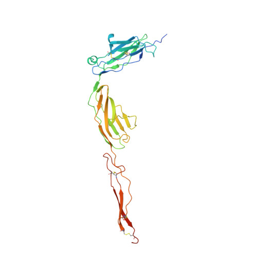
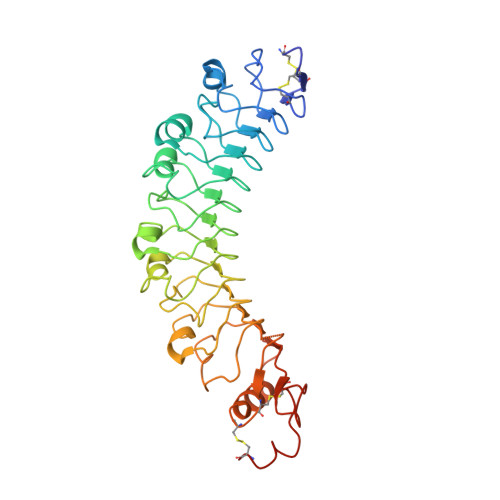
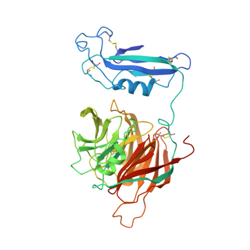
    Latrophilin adhesion-GPCRs (Lphn1-3 or ADGRL1-3) and Unc5 cell guidance receptors (Unc5A-D) interact with FLRT proteins (FLRT1-3), thereby promoting cell adhesion and repulsion, respectively. How the three proteins interact and function simultaneously is poorly understood. We show that Unc5D interacts with FLRT2 in cis, controlling cell adhesion in response to externally presented Lphn3. The ectodomains of the three proteins bind cooperatively. Crystal structures of the ternary complex formed by the extracellular domains reveal that Lphn3 dimerizes when bound to FLRT2:Unc5, resulting in a stoichiometry of 1:1:2 (FLRT2:Unc5D:Lphn3). This 1:1:2 complex further dimerizes to form a larger 'super-complex' (2:2:4), using a previously undescribed binding motif in the Unc5D TSP1 domain. Molecular dynamics simulations, point-directed mutagenesis and mass spectrometry demonstrate the stability and molecular properties of these complexes. Our data exemplify how receptors increase their functional repertoire by forming different context-dependent higher-order complexes.


  • Organizational Affiliation

    Department of Biochemistry, Oxford University, Oxford OX1 3QU, UK.


Macromolecules
Find similar proteins by:  (by identity cutoff)  |  3D Structure
Entity ID: 1
MoleculeChains Sequence LengthOrganismDetailsImage
NETRIN RECEPTOR UNC5D
A, E
285Rattus norvegicusMutation(s): 0 
UniProt
Find proteins for F1LW30 (Rattus norvegicus)
Explore F1LW30 
Go to UniProtKB:  F1LW30
Entity Groups  
Sequence Clusters30% Identity50% Identity70% Identity90% Identity95% Identity100% Identity
UniProt GroupF1LW30
Sequence Annotations
Expand
  • Reference Sequence
Find similar proteins by:  (by identity cutoff)  |  3D Structure
Entity ID: 2
MoleculeChains Sequence LengthOrganismDetailsImage
LEUCINE-RICH REPEAT TRANSMEMBRANE PROTEIN FLRT2
B, F
339Mus musculusMutation(s): 0 
UniProt
Find proteins for Q8BLU0 (Mus musculus)
Explore Q8BLU0 
Go to UniProtKB:  Q8BLU0
Entity Groups  
Sequence Clusters30% Identity50% Identity70% Identity90% Identity95% Identity100% Identity
UniProt GroupQ8BLU0
Sequence Annotations
Expand
  • Reference Sequence
Find similar proteins by:  (by identity cutoff)  |  3D Structure
Entity ID: 3
MoleculeChains Sequence LengthOrganismDetailsImage
ADHESION G PROTEIN-COUPLED RECEPTOR L3
C, D, G, H
383Mus musculusMutation(s): 0 
UniProt & NIH Common Fund Data Resources
Find proteins for Q80TS3 (Mus musculus)
Explore Q80TS3 
Go to UniProtKB:  Q80TS3
IMPC:  MGI:2441950
Entity Groups  
Sequence Clusters30% Identity50% Identity70% Identity90% Identity95% Identity100% Identity
UniProt GroupQ80TS3
Sequence Annotations
Expand
  • Reference Sequence
Small Molecules
Ligands 3 Unique
IDChains Name / Formula / InChI Key2D Diagram3D Interactions
NAG
Query on NAG

Download Ideal Coordinates CCD File 
I [auth A]
J [auth A]
K [auth B]
L [auth C]
Q [auth E]
I [auth A],
J [auth A],
K [auth B],
L [auth C],
Q [auth E],
R [auth E],
S [auth F],
T [auth G]
2-acetamido-2-deoxy-beta-D-glucopyranose
C8 H15 N O6
OVRNDRQMDRJTHS-FMDGEEDCSA-N
CA
Query on CA

Download Ideal Coordinates CCD File 
N [auth C],
P [auth D],
V [auth G],
X [auth H]
CALCIUM ION
Ca
BHPQYMZQTOCNFJ-UHFFFAOYSA-N
NA
Query on NA

Download Ideal Coordinates CCD File 
M [auth C],
O [auth D],
U [auth G],
W [auth H]
SODIUM ION
Na
FKNQFGJONOIPTF-UHFFFAOYSA-N
Experimental Data & Validation

Experimental Data

  • Method: X-RAY DIFFRACTION
  • Resolution: 3.40 Å
  • R-Value Free: 0.245 
  • R-Value Work: 0.225 
  • R-Value Observed: 0.226 
  • Space Group: C 1 2 1
Unit Cell:
Length ( Å )Angle ( ˚ )
a = 231.961α = 90
b = 141.486β = 117.94
c = 151.491γ = 90
Software Package:
Software NamePurpose
BUSTERrefinement
XDSdata reduction
Aimlessdata scaling
PHASERphasing

Structure Validation

View Full Validation Report


Ligand Structure Quality Assessment 


Entry History 

Revision History  (Full details and data files)

  • Version 1.0: 2016-05-04
    Type: Initial release
  • Version 1.1: 2020-07-29
    Type: Remediation
    Reason: Carbohydrate remediation
    Changes: Advisory, Data collection, Derived calculations, Other, Structure summary