5FRA

CBM40_CPF0721-6'SL


Experimental Data Snapshot

  • Method: X-RAY DIFFRACTION
  • Resolution: 2.00 Å
  • R-Value Free: 0.210 
  • R-Value Work: 0.179 
  • R-Value Observed: 0.180 

wwPDB Validation   3D Report Full Report


Ligand Structure Quality Assessment 


This is version 2.1 of the entry. See complete history


Literature

Characterization of a High-Affinity Sialic Acid-Specific Cbm40 from Clostridium Perfringens and Engineering of a Divalent Form.

Ribeiro, J.P.Pau, W.Pifferi, C.Renaudet, O.Varrot, A.Mahal, L.K.Imberty, A.

(2016) Biochem J 473: 2109

  • DOI: https://doi.org/10.1042/BCJ20160340
  • Primary Citation of Related Structures:  
    5FRA, 5FRE

  • PubMed Abstract: 

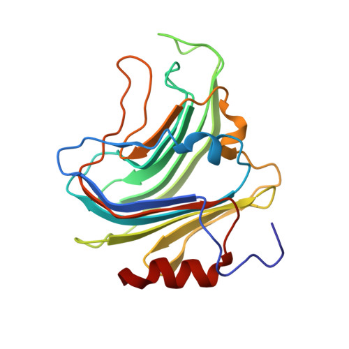
    CBMs (carbohydrate-binding modules) are a class of polypeptides usually associated with carbohydrate-active enzymatic sites. We have characterized a new member of the CBM40 family, coded from a section of the gene NanI from Clostridium perfringens Glycan arrays revealed its preference towards α(2,3)-linked sialosides, which was confirmed and quantified by calorimetric studies. The CBM40 binds to α(2,3)-sialyl-lactose with a Kd of ∼30 μM, the highest affinity value for this class of proteins. Inspired by lectins' structure and their arrangement as multimeric proteins, we have engineered a dimeric form of the CBM, and using SPR (surface plasmon resonance) we have observed 6-11-fold binding increases due to the avidity affect. The structures of the CBM, resolved by X-ray crystallography, in complex with α(2,3)- or α(2,6)-sialyl-lactose explain its binding specificity and unusually strong binding.


  • Organizational Affiliation

    Biomedical Chemistry Institute, New York University Department of Chemistry, 100 Washington Square East, Room 1001, New York, NY 10003, U.S.A. CERMAV, UPR5301, CNRS and Université Grenoble Alpes, 601 rue de la Chimie, BP 53, 38041, Grenoble, France.


Macromolecules
Find similar proteins by:  (by identity cutoff)  |  3D Structure
Entity ID: 1
MoleculeChains Sequence LengthOrganismDetailsImage
SIALIDASE
A, B, C, D, E
A, B, C, D, E, F
197Clostridium perfringensMutation(s): 0 
UniProt
Find proteins for A0A0H2YQR1 (Clostridium perfringens (strain ATCC 13124 / DSM 756 / JCM 1290 / NCIMB 6125 / NCTC 8237 / Type A))
Explore A0A0H2YQR1 
Go to UniProtKB:  A0A0H2YQR1
Entity Groups  
Sequence Clusters30% Identity50% Identity70% Identity90% Identity95% Identity100% Identity
UniProt GroupA0A0H2YQR1
Sequence Annotations
Expand
  • Reference Sequence
Oligosaccharides

Help

Entity ID: 2
MoleculeChains Length2D Diagram Glycosylation3D Interactions
N-acetyl-alpha-neuraminic acid-(2-6)-beta-D-galactopyranose
G, H, I
2N/A
Glycosylation Resources
GlyTouCan:  G63069TR
GlyCosmos:  G63069TR
GlyGen:  G63069TR
Experimental Data & Validation

Experimental Data

  • Method: X-RAY DIFFRACTION
  • Resolution: 2.00 Å
  • R-Value Free: 0.210 
  • R-Value Work: 0.179 
  • R-Value Observed: 0.180 
  • Space Group: P 1 21 1
Unit Cell:
Length ( Å )Angle ( ˚ )
a = 78.502α = 90
b = 84.267β = 90.79
c = 89.996γ = 90
Software Package:
Software NamePurpose
REFMACrefinement
XDSdata reduction
Aimlessdata scaling
PHASERphasing

Structure Validation

View Full Validation Report


Ligand Structure Quality Assessment 


Entry History 

Deposition Data

Revision History  (Full details and data files)

  • Version 1.0: 2016-07-20
    Type: Initial release
  • Version 1.1: 2017-09-27
    Changes: Data collection
  • Version 2.0: 2020-07-29
    Type: Remediation
    Reason: Carbohydrate remediation
    Changes: Advisory, Atomic model, Data collection, Derived calculations, Other, Structure summary
  • Version 2.1: 2024-01-10
    Changes: Data collection, Database references, Refinement description, Structure summary