5F8W

Crystal structure of a Crenomytilus grayanus lectin in complex with galactose


Experimental Data Snapshot

  • Method: X-RAY DIFFRACTION
  • Resolution: 1.56 Å
  • R-Value Free: 0.139 
  • R-Value Work: 0.105 
  • R-Value Observed: 0.107 

wwPDB Validation   3D Report Full Report


Ligand Structure Quality Assessment 


This is version 1.3 of the entry. See complete history


Literature

A Multivalent Marine Lectin from Crenomytilus grayanus Possesses Anti-cancer Activity through Recognizing Globotriose Gb3

Liao, J.-H.Chien, C.-T.Wu, H.-Y.Huang, K.-F.Wang, I.Ho, M.-R.Tu, I.-F.Lee, I.-M.Li, W.Shih, Y.-L.Wu, C.-Y.Lukyanov, P.A.Hsu, S.D.Wu, S.-H.

(2016) J Am Chem Soc 138: 4787-4795

  • DOI: https://doi.org/10.1021/jacs.6b00111
  • Primary Citation of Related Structures:  
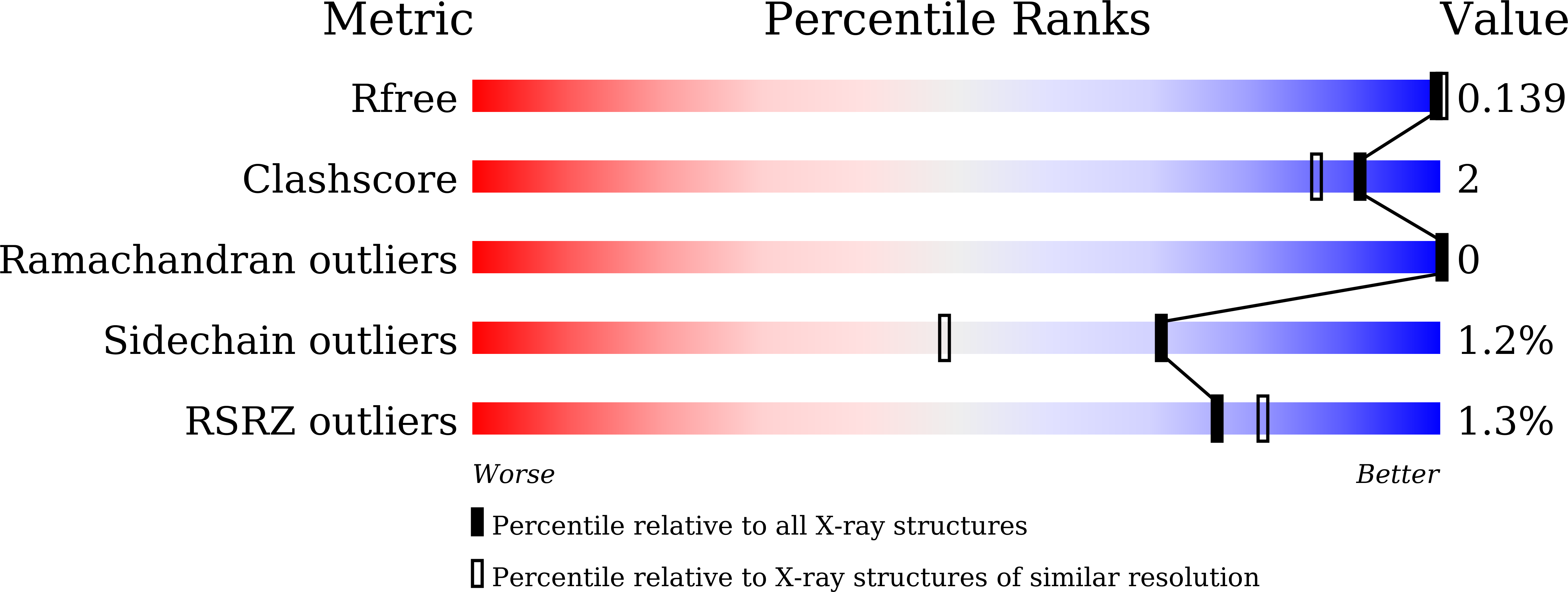
    5F8S, 5F8W, 5F8Y, 5F90

  • PubMed Abstract: 

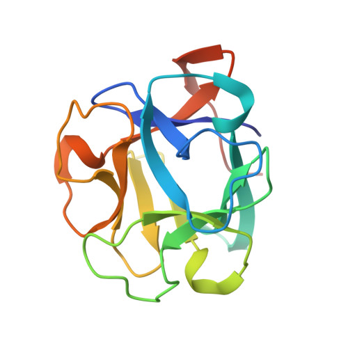
    In this study, we report the structure and function of a lectin from the sea mollusk Crenomytilus grayanus collected from the sublittoral zone of Peter the Great Bay of the Sea of Japan. The crystal structure of C. grayanus lectin (CGL) was solved to a resolution of 1.08 Å, revealing a β-trefoil fold that dimerizes into a dumbbell-shaped quaternary structure. Analysis of the crystal CGL structures bound to galactose, galactosamine, and globotriose Gb3 indicated that each CGL can bind three ligands through a carbohydrate-binding motif involving an extensive histidine- and water-mediated hydrogen bond network. CGL binding to Gb3 is further enhanced by additional side-chain-mediated hydrogen bonds in each of the three ligand-binding sites. NMR titrations revealed that the three binding sites have distinct microscopic affinities toward galactose and galactosamine. Cell viability assays showed that CGL recognizes Gb3 on the surface of breast cancer cells, leading to cell death. Our findings suggest the use of this lectin in cancer diagnosis and treatment.


  • Organizational Affiliation

    Institute of Biological Chemistry, Academia Sinica , Taipei 11529, Taiwan.


Macromolecules
Find similar proteins by:  (by identity cutoff)  |  3D Structure
Entity ID: 1
MoleculeChains Sequence LengthOrganismDetailsImage
GalNAc/Gal-specific lectin
A, B
156Crenomytilus grayanusMutation(s): 0 
UniProt
Find proteins for H2FH31 (Crenomytilus grayanus)
Explore H2FH31 
Go to UniProtKB:  H2FH31
Entity Groups  
Sequence Clusters30% Identity50% Identity70% Identity90% Identity95% Identity100% Identity
UniProt GroupH2FH31
Sequence Annotations
Expand
  • Reference Sequence
Small Molecules
Ligands 3 Unique
IDChains Name / Formula / InChI Key2D Diagram3D Interactions
GLA
Query on GLA

Download Ideal Coordinates CCD File 
D [auth A],
E [auth A],
F [auth B],
G [auth B],
H [auth B]
alpha-D-galactopyranose
C6 H12 O6
WQZGKKKJIJFFOK-PHYPRBDBSA-N
GAL
Query on GAL

Download Ideal Coordinates CCD File 
C [auth A]beta-D-galactopyranose
C6 H12 O6
WQZGKKKJIJFFOK-FPRJBGLDSA-N
GOL
Query on GOL

Download Ideal Coordinates CCD File 
I [auth B],
J [auth B]
GLYCEROL
C3 H8 O3
PEDCQBHIVMGVHV-UHFFFAOYSA-N
Binding Affinity Annotations 
IDSourceBinding Affinity
GAL Binding MOAD:  5F8W Kd: 1.50e+4 (nM) from 1 assay(s)
GLA Binding MOAD:  5F8W Kd: 5.10e+4 (nM) from 1 assay(s)
Experimental Data & Validation

Experimental Data

  • Method: X-RAY DIFFRACTION
  • Resolution: 1.56 Å
  • R-Value Free: 0.139 
  • R-Value Work: 0.105 
  • R-Value Observed: 0.107 
  • Space Group: P 21 21 21
Unit Cell:
Length ( Å )Angle ( ˚ )
a = 53.26α = 90
b = 72.542β = 90
c = 94.622γ = 90
Software Package:
Software NamePurpose
REFMACrefinement
HKL-2000data reduction
HKL-2000data scaling
SHELXCDphasing

Structure Validation

View Full Validation Report


Ligand Structure Quality Assessment 


Entry History & Funding Information

Deposition Data


Funding OrganizationLocationGrant Number
National Science Council and Ministry of Science and TechnologyTaiwanNSC 100-2113-M-001-022-MY3, NSC 102-2811-M-001-132, MOST 103-2113-M-001-029-MY3, MOST 103-2811-M-001-126, NSC 103-2923-M-001-006-MY3, NSC 100-2113-M-001-031-MY2, NSC 102-2113-M-001-017-MY2

Revision History  (Full details and data files)

  • Version 1.0: 2016-04-06
    Type: Initial release
  • Version 1.1: 2016-04-27
    Changes: Database references
  • Version 1.2: 2020-07-29
    Type: Remediation
    Reason: Carbohydrate remediation
    Changes: Data collection, Derived calculations, Structure summary
  • Version 1.3: 2024-03-20
    Changes: Data collection, Database references, Structure summary