


Crystal structure of Fructokinase from Vibrio cholerae O395 in apo form
Paul, R., Nath, S., Sen, U.To be published.
Experimental Data Snapshot
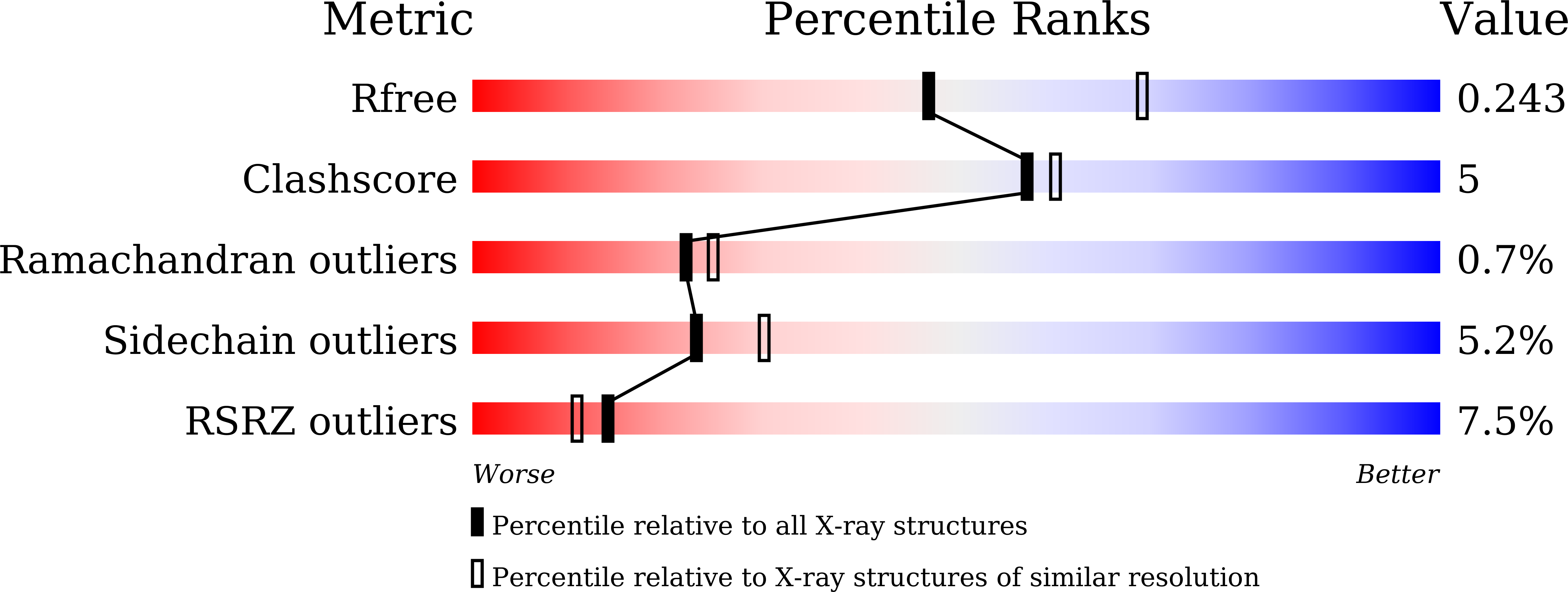
wwPDB Validation 3D Report Full Report
Entity ID: 1 | |||||
|---|---|---|---|---|---|
| Molecule | Chains | Sequence Length | Organism | Details | Image |
| Fructokinase | 323 | Vibrio cholerae O395 | Mutation(s): 0 Gene Names: cscK, VC0395_0600 EC: 2.7.1.4 | 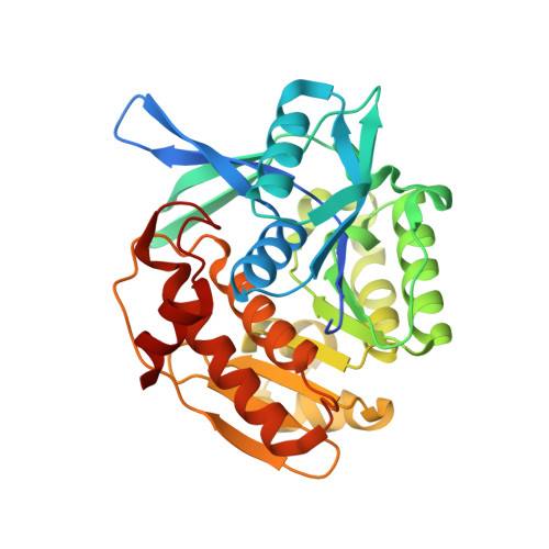 | |
UniProt | |||||
Find proteins for A0A0H3AER7 (Vibrio cholerae serotype O1 (strain ATCC 39541 / Classical Ogawa 395 / O395)) Explore A0A0H3AER7 Go to UniProtKB: A0A0H3AER7 | |||||
Entity Groups | |||||
| Sequence Clusters | 30% Identity50% Identity70% Identity90% Identity95% Identity100% Identity | ||||
| UniProt Group | A0A0H3AER7 | ||||
Sequence AnnotationsExpand | |||||
| |||||
| Ligands 1 Unique | |||||
|---|---|---|---|---|---|
| ID | Chains | Name / Formula / InChI Key | 2D Diagram | 3D Interactions | |
| NA Query on NA | C [auth A] | SODIUM ION Na FKNQFGJONOIPTF-UHFFFAOYSA-N |  | ||
| Length ( Å ) | Angle ( ˚ ) |
|---|---|
| a = 107.116 | α = 90 |
| b = 99.858 | β = 90 |
| c = 61.548 | γ = 90 |
| Software Name | Purpose |
|---|---|
| PHENIX | refinement |
| AUTOMAR | data reduction |
| AUTOMAR | data scaling |
| MOLREP | phasing |
RCSB PDB (citation) is hosted by
RCSB PDB is a member of the

