5EWR

C merolae U4 snRNP protein Snu13


Experimental Data Snapshot

  • Method: X-RAY DIFFRACTION
  • Resolution: 2.35 Å
  • R-Value Free: 0.220 
  • R-Value Work: 0.166 
  • R-Value Observed: 0.169 

wwPDB Validation   3D Report Full Report


This is version 1.1 of the entry. See complete history


Literature

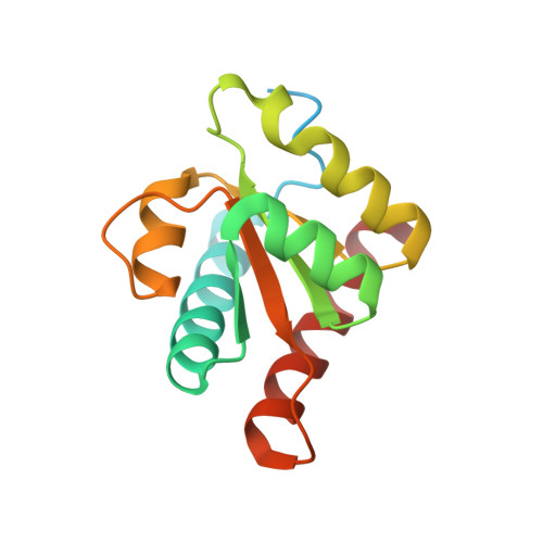
Conserved structure of Snu13 from the highly reduced spliceosome of Cyanidioschyzon merolae.

Black, C.S.Garside, E.L.MacMillan, A.M.Rader, S.D.

(2016) Protein Sci 25: 911-916

  • DOI: https://doi.org/10.1002/pro.2894
  • Primary Citation of Related Structures:  
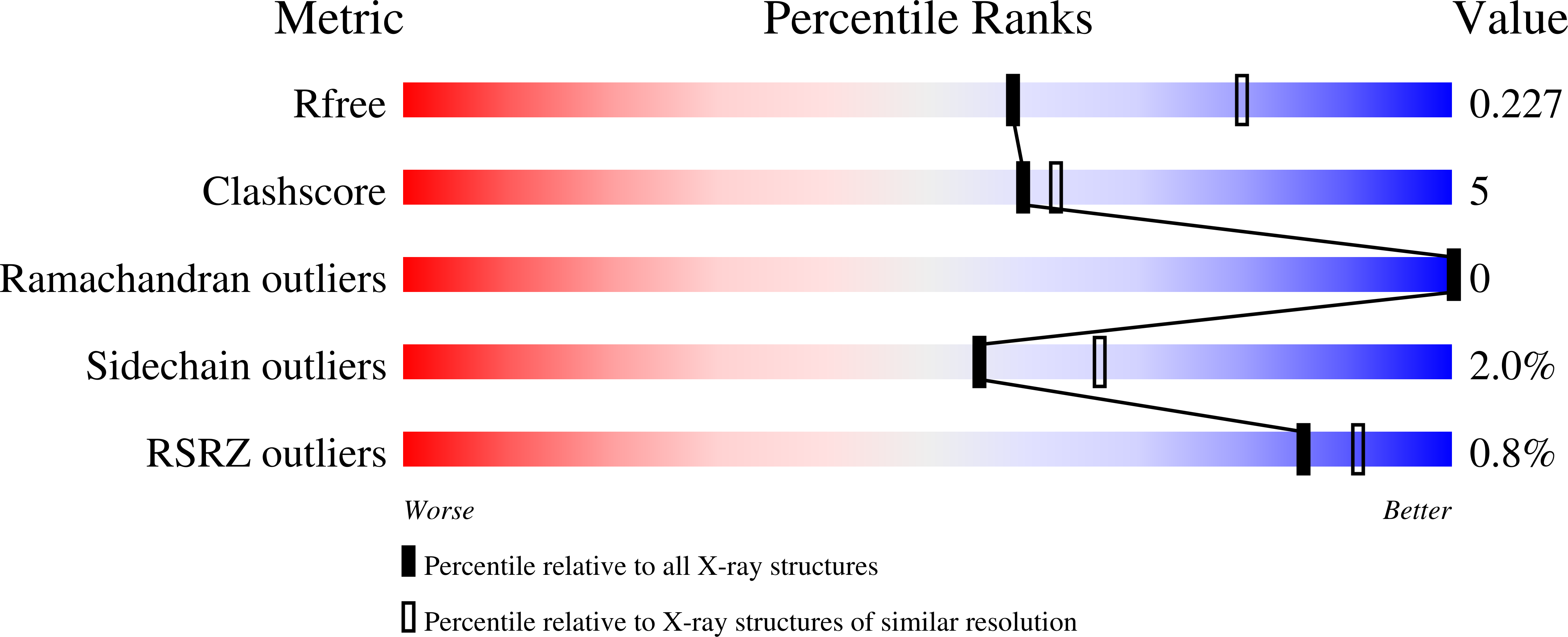
    5EWR

  • PubMed Abstract: 

    Structural and functional analysis of proteins involved in pre-mRNA splicing is challenging because of the complexity of the splicing machinery, known as the spliceosome. Bioinformatic, proteomic, and biochemical analyses have identified a minimal spliceosome in the red alga Cyanidioschyzon merolae. This spliceosome consists of only 40 core proteins, compared to ∼ 70 in S. cerevisiae (yeast) and ∼ 150 in humans. We report the X-ray crystallographic analysis of C. merolae Snu13 (CmSnu13), a key component of the assembling spliceosome, and present evidence for conservation of Snu13 function in this algal splicing pathway. The near identity of CmSnu13's three-dimensional structure to yeast and human Snu13 suggests that C. merolae should be an excellent model system for investigating the structure and function of the conserved core of the spliceosome.


  • Organizational Affiliation

    Department of Chemistry, University of Northern British Columbia, Prince George, British Columbia, V2N 4Z9, Canada.


Macromolecules
Find similar proteins by:  (by identity cutoff)  |  3D Structure
Entity ID: 1
MoleculeChains Sequence LengthOrganismDetailsImage
Box C/D snoRNP and U4 snRNP component Snu13p146Cyanidioschyzon merolaeMutation(s): 0 
Gene Names: CYME_CMP335C
UniProt
Find proteins for M1VFX0 (Cyanidioschyzon merolae (strain NIES-3377 / 10D))
Explore M1VFX0 
Go to UniProtKB:  M1VFX0
Entity Groups  
Sequence Clusters30% Identity50% Identity70% Identity90% Identity95% Identity100% Identity
UniProt GroupM1VFX0
Sequence Annotations
Expand
  • Reference Sequence
Experimental Data & Validation

Experimental Data

  • Method: X-RAY DIFFRACTION
  • Resolution: 2.35 Å
  • R-Value Free: 0.220 
  • R-Value Work: 0.166 
  • R-Value Observed: 0.169 
  • Space Group: P 21 21 21
Unit Cell:
Length ( Å )Angle ( ˚ )
a = 30.33α = 90
b = 57.582β = 90
c = 65.38γ = 90
Software Package:
Software NamePurpose
PHENIXrefinement
HKL-2000data collection
HKL-2000data scaling
PHASERphasing
PDB_EXTRACTdata extraction
HKL-2000data reduction

Structure Validation

View Full Validation Report


Entry History 

Deposition Data

Revision History  (Full details and data files)

  • Version 1.0: 2016-04-20
    Type: Initial release
  • Version 1.1: 2023-09-27
    Changes: Data collection, Database references, Derived calculations, Refinement description