5ERQ

Gephyrin E domain at 1.55 angstrom resolution


Experimental Data Snapshot

  • Method: X-RAY DIFFRACTION
  • Resolution: 1.55 Å
  • R-Value Free: 0.187 
  • R-Value Work: 0.154 
  • R-Value Observed: 0.156 

wwPDB Validation   3D Report Full Report


This is version 2.0 of the entry. See complete history


Literature

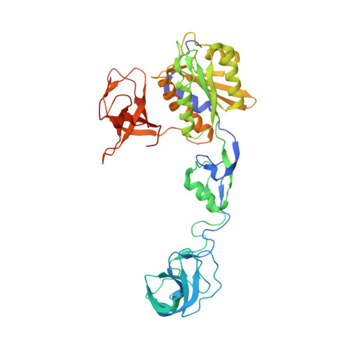
Structural Framework for Metal Incorporation during Molybdenum Cofactor Biosynthesis.

Kasaragod, V.B.Schindelin, H.

(2016) Structure 24: 782-788

  • DOI: https://doi.org/10.1016/j.str.2016.02.023
  • Primary Citation of Related Structures:  
    5ERQ, 5ERR, 5ERS, 5ERT, 5ERU, 5ERV

  • PubMed Abstract: 

    The molybdenum cofactor (Moco) is essential for the catalytic activity of all molybdenum-containing enzymes with the exception of nitrogenase. Moco biosynthesis follows an evolutionarily highly conserved pathway and genetic deficiencies in the corresponding human enzymes result in Moco deficiency, which manifests itself in severe neurological symptoms and death in childhood. In humans the final steps of Moco biosynthesis are catalyzed by gephyrin, specifically the penultimate adenylation of molybdopterin (MPT) by its N-terminal G domain (GephG) and the final metal incorporation by its C-terminal E domain (GephE). To better understand the poorly defined molecular framework of this final step, we determined high-resolution crystal structures of GephE in the apo state and in complex with ADP, AMP, and molybdate. Our data provide novel insights into the catalytic steps leading to final Moco maturation, namely deadenylation as well as molybdate binding and insertion.


  • Organizational Affiliation

    Rudolf Virchow Center for Experimental Biomedicine, Institute of Structural Biology, University of Würzburg, Josef-Schneider-Straße 2, 97080 Würzburg, Germany.


Macromolecules
Find similar proteins by:  (by identity cutoff)  |  3D Structure
Entity ID: 1
MoleculeChains Sequence LengthOrganismDetailsImage
Gephyrin419Rattus norvegicusMutation(s): 0 
Gene Names: GphnGph
EC: 2.7.7.75 (PDB Primary Data), 2.10.1.1 (PDB Primary Data)
UniProt
Find proteins for Q03555 (Rattus norvegicus)
Explore Q03555 
Go to UniProtKB:  Q03555
Entity Groups  
Sequence Clusters30% Identity50% Identity70% Identity90% Identity95% Identity100% Identity
UniProt GroupQ03555
Sequence Annotations
Expand
  • Reference Sequence
Small Molecules
Ligands 2 Unique
IDChains Name / Formula / InChI Key2D Diagram3D Interactions
MPD
Query on MPD

Download Ideal Coordinates CCD File 
L [auth A],
M [auth A]
(4S)-2-METHYL-2,4-PENTANEDIOL
C6 H14 O2
SVTBMSDMJJWYQN-YFKPBYRVSA-N
ACT
Query on ACT

Download Ideal Coordinates CCD File 
B [auth A]
C [auth A]
D [auth A]
E [auth A]
F [auth A]
B [auth A],
C [auth A],
D [auth A],
E [auth A],
F [auth A],
G [auth A],
H [auth A],
I [auth A],
J [auth A],
K [auth A]
ACETATE ION
C2 H3 O2
QTBSBXVTEAMEQO-UHFFFAOYSA-M
Experimental Data & Validation

Experimental Data

  • Method: X-RAY DIFFRACTION
  • Resolution: 1.55 Å
  • R-Value Free: 0.187 
  • R-Value Work: 0.154 
  • R-Value Observed: 0.156 
  • Space Group: I 2 2 2
Unit Cell:
Length ( Å )Angle ( ˚ )
a = 87.669α = 90
b = 99.287β = 90
c = 113.725γ = 90
Software Package:
Software NamePurpose
PHENIXrefinement
XDSdata reduction
XDSdata scaling
PHASERphasing

Structure Validation

View Full Validation Report


Entry History 

Deposition Data

Revision History  (Full details and data files)

  • Version 1.0: 2016-05-04
    Type: Initial release
  • Version 1.1: 2016-05-11
    Changes: Database references
  • Version 2.0: 2024-01-10
    Changes: Atomic model, Data collection, Database references, Refinement description