


Crystal structure of NAD kinase 1 from Listeria monocytogenes: P252D mutant
Poncet-Montange, G., Assairi, L., Gelin, M., Pochet, S., Labesse, G.To be published.
Experimental Data Snapshot
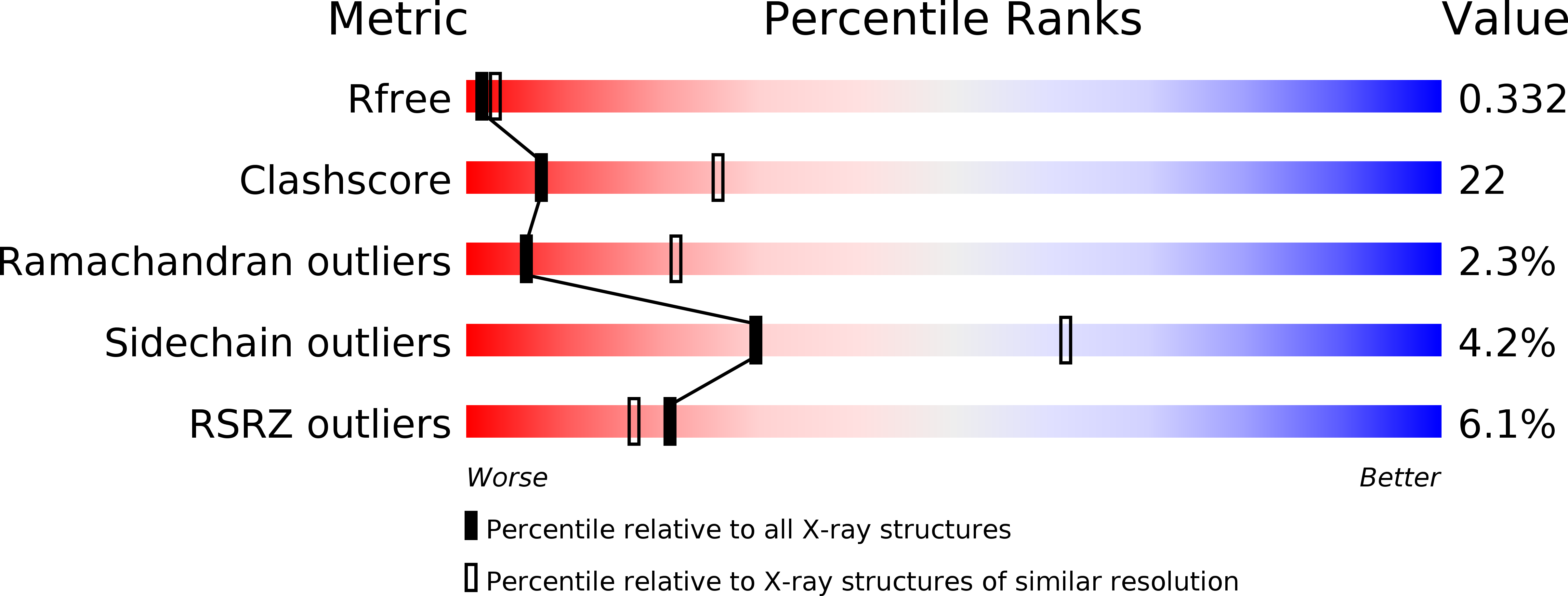
wwPDB Validation 3D Report Full Report
Entity ID: 1 | |||||
|---|---|---|---|---|---|
| Molecule | Chains | Sequence Length | Organism | Details | Image |
| NAD kinase 1 | 272 | Listeria monocytogenes | Mutation(s): 1 Gene Names: nadK1, lmo0968 EC: 2.7.1.23 | 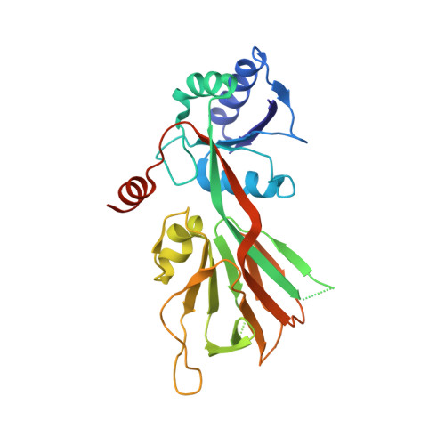 | |
UniProt | |||||
Find proteins for Q8Y8D7 (Listeria monocytogenes serovar 1/2a (strain ATCC BAA-679 / EGD-e)) Explore Q8Y8D7 Go to UniProtKB: Q8Y8D7 | |||||
Entity Groups | |||||
| Sequence Clusters | 30% Identity50% Identity70% Identity90% Identity95% Identity100% Identity | ||||
| UniProt Group | Q8Y8D7 | ||||
Sequence AnnotationsExpand | |||||
| |||||
| Length ( Å ) | Angle ( ˚ ) |
|---|---|
| a = 67.217 | α = 90 |
| b = 119.655 | β = 100.92 |
| c = 68.624 | γ = 90 |
| Software Name | Purpose |
|---|---|
| PHENIX | refinement |
| SCALA | data scaling |
| PDB_EXTRACT | data extraction |
| Funding Organization | Location | Grant Number |
|---|---|---|
| French National Research Agency | France | -- |
RCSB PDB (citation) is hosted by
RCSB PDB is a member of the

