5EFV

The host-recognition device of Staphylococcus aureus phage Phi11


Experimental Data Snapshot

  • Method: X-RAY DIFFRACTION
  • Resolution: 2.20 Å
  • R-Value Free: 0.211 
  • R-Value Work: 0.173 
  • R-Value Observed: 0.175 

wwPDB Validation   3D Report Full Report


This is version 1.1 of the entry. See complete history


Literature

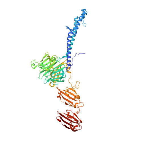
Structure of the host-recognition device of Staphylococcus aureus phage 11.

Koc, C.Xia, G.Kuhner, P.Spinelli, S.Roussel, A.Cambillau, C.Stehle, T.

(2016) Sci Rep 6: 27581-27581

  • DOI: https://doi.org/10.1038/srep27581
  • Primary Citation of Related Structures:  
    5EFV

  • PubMed Abstract: 

    Phages play key roles in the pathogenicity and adaptation of the human pathogen Staphylococcus aureus. However, little is known about the molecular recognition events that mediate phage adsorption to the surface of S. aureus. The lysogenic siphophage ϕ11 infects S. aureus SA113. It was shown previously that ϕ11 requires α- or β-N-acetylglucosamine (GlcNAc) moieties on cell wall teichoic acid (WTA) for adsorption. Gp45 was identified as the receptor binding protein (RBP) involved in this process and GlcNAc residues on WTA were found to be the key component of the ϕ11 receptor. Here we report the crystal structure of the RBP of ϕ11, which assembles into a large, multidomain homotrimer. Each monomer contains a five-bladed propeller domain with a cavity that could accommodate a GlcNAc moiety. An electron microscopy reconstruction of the ϕ11 host adhesion component, the baseplate, reveals that six RBP trimers are assembled around the baseplate core. The Gp45 and baseplate structures provide insights into the overall organization and molecular recognition process of the phage ϕ11 tail. This assembly is conserved among most glycan-recognizing Siphoviridae, and the RBP orientation would allow host adhesion and infection without an activation step.


  • Organizational Affiliation

    Interfaculty Institute of Biochemistry, University of Tübingen, 72076 Tübingen, Germany.


Macromolecules
Find similar proteins by:  (by identity cutoff)  |  3D Structure
Entity ID: 1
MoleculeChains Sequence LengthOrganismDetailsImage
Phi ETA orf 56-like protein
A, B, C
648Dubowvirus dv11Mutation(s): 0 
UniProt
Find proteins for Q8SDT4 (Staphylococcus phage phi 11)
Explore Q8SDT4 
Go to UniProtKB:  Q8SDT4
Entity Groups  
Sequence Clusters30% Identity50% Identity70% Identity90% Identity95% Identity100% Identity
UniProt GroupQ8SDT4
Sequence Annotations
Expand
  • Reference Sequence
Small Molecules
Ligands 1 Unique
IDChains Name / Formula / InChI Key2D Diagram3D Interactions
FE
Query on FE

Download Ideal Coordinates CCD File 
D [auth A]FE (III) ION
Fe
VTLYFUHAOXGGBS-UHFFFAOYSA-N
Experimental Data & Validation

Experimental Data

  • Method: X-RAY DIFFRACTION
  • Resolution: 2.20 Å
  • R-Value Free: 0.211 
  • R-Value Work: 0.173 
  • R-Value Observed: 0.175 
  • Space Group: P 1
Unit Cell:
Length ( Å )Angle ( ˚ )
a = 87.06α = 93.02
b = 89.01β = 105.25
c = 93.26γ = 117.58
Software Package:
Software NamePurpose
BUSTERrefinement
XDSdata reduction
XDSdata scaling
SHARPphasing

Structure Validation

View Full Validation Report


Entry History & Funding Information

Deposition Data


Funding OrganizationLocationGrant Number
Germany--

Revision History  (Full details and data files)

  • Version 1.0: 2016-05-25
    Type: Initial release
  • Version 1.1: 2016-06-22
    Changes: Database references