5DZX

Protocadherin beta 6 extracellular cadherin domains 1-4


Experimental Data Snapshot

  • Method: X-RAY DIFFRACTION
  • Resolution: 2.88 Å
  • R-Value Free: 0.289 
  • R-Value Work: 0.241 
  • R-Value Observed: 0.244 

wwPDB Validation   3D Report Full Report


Ligand Structure Quality Assessment 


This is version 2.1 of the entry. See complete history


Literature

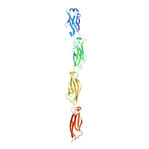
Structural Basis of Diverse Homophilic Recognition by Clustered alpha- and beta-Protocadherins.

Goodman, K.M.Rubinstein, R.Thu, C.A.Bahna, F.Mannepalli, S.Ahlsen, G.Rittenhouse, C.Maniatis, T.Honig, B.Shapiro, L.

(2016) Neuron 90: 709-723

  • DOI: https://doi.org/10.1016/j.neuron.2016.04.004
  • Primary Citation of Related Structures:  
    5DZV, 5DZW, 5DZX, 5DZY

  • PubMed Abstract: 

    Clustered protocadherin proteins (α-, β-, and γ-Pcdhs) provide a high level of cell-surface diversity to individual vertebrate neurons, engaging in highly specific homophilic interactions to mediate important roles in mammalian neural circuit development. How Pcdhs bind homophilically through their extracellular cadherin (EC) domains among dozens of highly similar isoforms has not been determined. Here, we report crystal structures for extracellular regions from four mouse Pcdh isoforms (α4, α7, β6, and β8), revealing a canonical head-to-tail interaction mode for homophilic trans dimers comprising primary intermolecular EC1:EC4 and EC2:EC3 interactions. A subset of trans interface residues exhibit isoform-specific conservation, suggesting roles in recognition specificity. Mutation of these residues, along with trans-interacting partner residues, altered the specificities of Pcdh interactions. Together, these data show how sequence variation among Pcdh isoforms encodes their diverse strict homophilic recognition specificities, which are required for their key roles in neural circuit assembly.


  • Organizational Affiliation

    Department of Biochemistry and Molecular Biophysics, Columbia University, New York, NY 10032, USA.


Macromolecules
Find similar proteins by:  (by identity cutoff)  |  3D Structure
Entity ID: 1
MoleculeChains Sequence LengthOrganismDetailsImage
Protocadherin beta 6
A, B
425Mus musculusMutation(s): 0 
Gene Names: Pcdhb6
UniProt
Find proteins for Q91XZ4 (Mus musculus)
Explore Q91XZ4 
Go to UniProtKB:  Q91XZ4
Entity Groups  
Sequence Clusters30% Identity50% Identity70% Identity90% Identity95% Identity100% Identity
UniProt GroupQ91XZ4
Sequence Annotations
Expand
  • Reference Sequence
Oligosaccharides

Help

Entity ID: 2
MoleculeChains Length2D Diagram Glycosylation3D Interactions
alpha-D-mannopyranose-(1-6)-beta-D-mannopyranose-(1-4)-2-acetamido-2-deoxy-beta-D-glucopyranose-(1-4)-[alpha-L-fucopyranose-(1-6)]2-acetamido-2-deoxy-beta-D-glucopyranose
C, D
5N-Glycosylation
Glycosylation Resources
GlyTouCan:  G00395TQ
GlyCosmos:  G00395TQ
GlyGen:  G00395TQ
Entity ID: 3
MoleculeChains Length2D Diagram Glycosylation3D Interactions
2-acetamido-2-deoxy-beta-D-glucopyranose-(1-4)-2-acetamido-2-deoxy-beta-D-glucopyranose
E
2N-Glycosylation
Glycosylation Resources
GlyTouCan:  G42666HT
GlyCosmos:  G42666HT
GlyGen:  G42666HT
Small Molecules
Ligands 2 Unique
IDChains Name / Formula / InChI Key2D Diagram3D Interactions
MAN
Query on MAN

Download Ideal Coordinates CCD File 
F [auth A],
P [auth B],
Q [auth B]
alpha-D-mannopyranose
C6 H12 O6
WQZGKKKJIJFFOK-PQMKYFCFSA-N
CA
Query on CA

Download Ideal Coordinates CCD File 
G [auth A]
H [auth A]
I [auth A]
J [auth A]
K [auth A]
G [auth A],
H [auth A],
I [auth A],
J [auth A],
K [auth A],
L [auth A],
M [auth A],
N [auth A],
O [auth A],
R [auth B],
S [auth B],
T [auth B],
U [auth B],
V [auth B],
W [auth B],
X [auth B],
Y [auth B],
Z [auth B]
CALCIUM ION
Ca
BHPQYMZQTOCNFJ-UHFFFAOYSA-N
Experimental Data & Validation

Experimental Data

  • Method: X-RAY DIFFRACTION
  • Resolution: 2.88 Å
  • R-Value Free: 0.289 
  • R-Value Work: 0.241 
  • R-Value Observed: 0.244 
  • Space Group: P 21 21 21
Unit Cell:
Length ( Å )Angle ( ˚ )
a = 49.04α = 90
b = 90.53β = 90
c = 258.13γ = 90
Software Package:
Software NamePurpose
PHENIXrefinement
XDSdata reduction
Aimlessdata scaling
PHASERphasing

Structure Validation

View Full Validation Report


Ligand Structure Quality Assessment 


Entry History 

Deposition Data

Revision History  (Full details and data files)

  • Version 1.0: 2016-05-04
    Type: Initial release
  • Version 1.1: 2016-05-25
    Changes: Database references
  • Version 1.2: 2016-06-01
    Changes: Database references
  • Version 2.0: 2020-07-29
    Type: Remediation
    Reason: Carbohydrate remediation
    Changes: Atomic model, Data collection, Database references, Derived calculations, Structure summary
  • Version 2.1: 2023-09-27
    Changes: Data collection, Database references, Derived calculations, Refinement description, Structure summary