5DUR

Influenza A virus H5 hemagglutinin globular head in complex with antibody 100F4


Experimental Data Snapshot

  • Method: X-RAY DIFFRACTION
  • Resolution: 2.82 Å
  • R-Value Free: 0.257 
  • R-Value Work: 0.208 
  • R-Value Observed: 0.210 

wwPDB Validation   3D Report Full Report


Ligand Structure Quality Assessment 


This is version 1.1 of the entry. See complete history


Literature

Comprehensive analysis of antibody recognition in convalescent humans from highly pathogenic avian influenza H5N1 infection

Zuo, T.Sun, J.Wang, G.Jiang, L.Zuo, Y.Li, D.Shi, X.Liu, X.Fan, S.Ren, H.Hu, H.Sun, L.Zhou, B.Liang, M.Zhou, P.Wang, X.Zhang, L.

(2015) Nat Commun 6: 8855-8855

  • DOI: https://doi.org/10.1038/ncomms9855
  • Primary Citation of Related Structures:  
    5DUM, 5DUP, 5DUR, 5DUT

  • PubMed Abstract: 

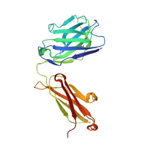
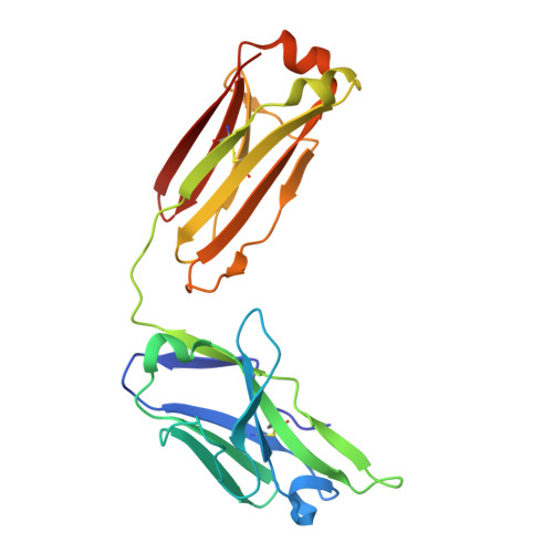
    Understanding the mechanism of protective antibody recognition against highly pathogenic avian influenza A virus H5N1 in humans is critical for the development of effective therapies and vaccines. Here we report the crystal structure of three H5-specific human monoclonal antibodies bound to the globular head of hemagglutinin (HA) with distinct epitope specificities, neutralization potencies and breadth. A structural and functional analysis of these epitopes combined with those reported elsewhere identifies four major vulnerable sites on the globular head of H5N1 HA. Chimeric and vulnerable site-specific mutant pseudoviruses are generated to delineate broad neutralization specificities of convalescent sera from two individuals who recovered from the infection with H5N1 virus. Our results show that the four vulnerable sites on the globular head rather than the stem region are the major neutralizing targets, suggesting that during natural H5N1 infection neutralizing antibodies against the globular head work in concert to provide protective antibody-mediated immunity.


  • Organizational Affiliation

    Comprehensive AIDS Research Center, Collaborative Innovation Center for Diagnosis and Treatment of Infectious Diseases, School of Medicine, Tsinghua University, Beijing 100084, China.


Macromolecules
Find similar proteins by:  (by identity cutoff)  |  3D Structure
Entity ID: 1
MoleculeChains Sequence LengthOrganismDetailsImage
Heavy Chain of Antibody 100F4A [auth H],
D [auth B]
231Homo sapiensMutation(s): 0 
Entity Groups  
Sequence Clusters30% Identity50% Identity70% Identity90% Identity95% Identity100% Identity
Sequence Annotations
Expand
  • Reference Sequence
Find similar proteins by:  (by identity cutoff)  |  3D Structure
Entity ID: 2
MoleculeChains Sequence LengthOrganismDetailsImage
Light Chain of Antibody 100F4B [auth L],
E [auth D]
218Homo sapiensMutation(s): 0 
Entity Groups  
Sequence Clusters30% Identity50% Identity70% Identity90% Identity95% Identity100% Identity
Sequence Annotations
Expand
  • Reference Sequence
Find similar proteins by:  (by identity cutoff)  |  3D Structure
Entity ID: 3
MoleculeChains Sequence LengthOrganismDetailsImage
HemagglutininC,
F [auth A]
233Influenza A virus (A/Anhui/1/2005(H5N1))Mutation(s): 0 
Gene Names: HA
UniProt
Find proteins for Q1WDM0 (Influenza A virus)
Explore Q1WDM0 
Go to UniProtKB:  Q1WDM0
Entity Groups  
Sequence Clusters30% Identity50% Identity70% Identity90% Identity95% Identity100% Identity
UniProt GroupQ1WDM0
Sequence Annotations
Expand
  • Reference Sequence
Small Molecules
Ligands 1 Unique
IDChains Name / Formula / InChI Key2D Diagram3D Interactions
NAG
Query on NAG

Download Ideal Coordinates CCD File 
G [auth A],
H [auth A]
2-acetamido-2-deoxy-beta-D-glucopyranose
C8 H15 N O6
OVRNDRQMDRJTHS-FMDGEEDCSA-N
Experimental Data & Validation

Experimental Data

  • Method: X-RAY DIFFRACTION
  • Resolution: 2.82 Å
  • R-Value Free: 0.257 
  • R-Value Work: 0.208 
  • R-Value Observed: 0.210 
  • Space Group: P 21 21 21
Unit Cell:
Length ( Å )Angle ( ˚ )
a = 88.094α = 90
b = 101.223β = 90
c = 206.301γ = 90
Software Package:
Software NamePurpose
PHENIXrefinement
HKL-2000data reduction
HKL-2000data scaling
PHASERphasing

Structure Validation

View Full Validation Report


Ligand Structure Quality Assessment 


Entry History 

Deposition Data

Revision History  (Full details and data files)

  • Version 1.0: 2015-12-16
    Type: Initial release
  • Version 1.1: 2020-07-29
    Type: Remediation
    Reason: Carbohydrate remediation
    Changes: Data collection, Derived calculations, Structure summary