5DMQ

Crystal structure of mouse eRF1 in complex with Reverse Transcriptase (RT) of Moloney Murine Leukemia Virus


Experimental Data Snapshot

  • Method: X-RAY DIFFRACTION
  • Resolution: 4.00 Å
  • R-Value Free: 0.295 
  • R-Value Work: 0.241 
  • R-Value Observed: 0.244 

wwPDB Validation   3D Report Full Report


This is version 1.2 of the entry. See complete history


Literature

Structural basis of suppression of host translation termination by Moloney Murine Leukemia Virus

Tang, X.Zhu, Y.Baker, S.L.Bowler, M.W.Chen, B.J.Chen, C.Hogg, J.R.Goff, S.P.Song, H.

(2016) Nat Commun 7: 12070-12070

  • DOI: https://doi.org/10.1038/ncomms12070
  • Primary Citation of Related Structures:  
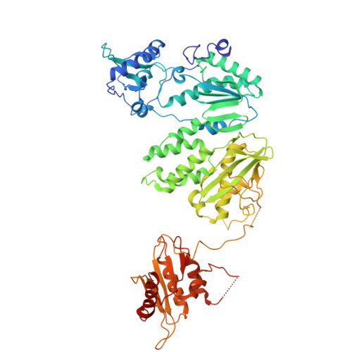
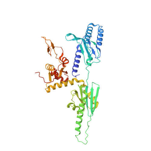
    5DMQ, 5DMR

  • PubMed Abstract: 

    Retroviral reverse transcriptase (RT) of Moloney murine leukemia virus (MoMLV) is expressed in the form of a large Gag-Pol precursor protein by suppression of translational termination in which the maximal efficiency of stop codon read-through depends on the interaction between MoMLV RT and peptidyl release factor 1 (eRF1). Here, we report the crystal structure of MoMLV RT in complex with eRF1. The MoMLV RT interacts with the C-terminal domain of eRF1 via its RNase H domain to sterically occlude the binding of peptidyl release factor 3 (eRF3) to eRF1. Promotion of read-through by MoMLV RNase H prevents nonsense-mediated mRNA decay (NMD) of mRNAs. Comparison of our structure with that of HIV RT explains why HIV RT cannot interact with eRF1. Our results provide a mechanistic view of how MoMLV manipulates the host translation termination machinery for the synthesis of its own proteins.


  • Organizational Affiliation

    Institute of Molecular and Cell Biology, 61 Biopolis Drive, Proteos, Singapore 138673, Singapore.


Macromolecules
Find similar proteins by:  (by identity cutoff)  |  3D Structure
Entity ID: 1
MoleculeChains Sequence LengthOrganismDetailsImage
Reverse transcriptase/ribonuclease H p80648Moloney murine leukemia virus isolate ShinnickMutation(s): 0 
EC: 2.7.7.49 (PDB Primary Data), 2.7.7.7 (PDB Primary Data), 3.1.26.4 (PDB Primary Data)
UniProt
Find proteins for P03355 (Moloney murine leukemia virus (isolate Shinnick))
Explore P03355 
Go to UniProtKB:  P03355
Entity Groups  
Sequence Clusters30% Identity50% Identity70% Identity90% Identity95% Identity100% Identity
UniProt GroupP03355
Sequence Annotations
Expand
  • Reference Sequence
Find similar proteins by:  (by identity cutoff)  |  3D Structure
Entity ID: 2
MoleculeChains Sequence LengthOrganismDetailsImage
Eukaryotic peptide chain release factor subunit 1437Mus musculusMutation(s): 0 
Gene Names: Etf1
UniProt
Find proteins for Q8BWY3 (Mus musculus)
Explore Q8BWY3 
Go to UniProtKB:  Q8BWY3
Entity Groups  
Sequence Clusters30% Identity50% Identity70% Identity90% Identity95% Identity100% Identity
UniProt GroupQ8BWY3
Sequence Annotations
Expand
  • Reference Sequence
Experimental Data & Validation

Experimental Data

  • Method: X-RAY DIFFRACTION
  • Resolution: 4.00 Å
  • R-Value Free: 0.295 
  • R-Value Work: 0.241 
  • R-Value Observed: 0.244 
  • Space Group: P 32 2 1
Unit Cell:
Length ( Å )Angle ( ˚ )
a = 92.653α = 90
b = 92.653β = 90
c = 289.949γ = 120
Software Package:
Software NamePurpose
REFMACrefinement
PHASERphasing
Cootmodel building

Structure Validation

View Full Validation Report


Entry History 

Deposition Data

Revision History  (Full details and data files)

  • Version 1.0: 2016-07-06
    Type: Initial release
  • Version 1.1: 2017-10-04
    Changes: Data collection, Derived calculations
  • Version 1.2: 2024-03-20
    Changes: Data collection, Database references