


Structure of mutant toxin
Parker, M.W., Gorman, M.A., Lawrence, S.L., Morton, C.J.To be published.
Experimental Data Snapshot
wwPDB Validation 3D Report Full Report

Entity ID: 1 | |||||
|---|---|---|---|---|---|
| Molecule | Chains | Sequence Length | Organism | Details | Image |
| Perfringolysin O | 472 | Clostridium perfringens str. 13 | Mutation(s): 1 Gene Names: pfo, pfoA, pfoR, CPE0163 | 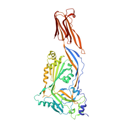 | |
UniProt | |||||
Find proteins for P0C2E9 (Clostridium perfringens (strain 13 / Type A)) Explore P0C2E9 Go to UniProtKB: P0C2E9 | |||||
Entity Groups | |||||
| Sequence Clusters | 30% Identity50% Identity70% Identity90% Identity95% Identity100% Identity | ||||
| UniProt Group | P0C2E9 | ||||
Sequence AnnotationsExpand | |||||
| |||||
| Ligands 1 Unique | |||||
|---|---|---|---|---|---|
| ID | Chains | Name / Formula / InChI Key | 2D Diagram | 3D Interactions | |
| EPE Query on EPE | C [auth A], D [auth B] | 4-(2-HYDROXYETHYL)-1-PIPERAZINE ETHANESULFONIC ACID C8 H18 N2 O4 S JKMHFZQWWAIEOD-UHFFFAOYSA-N |  | ||
| Length ( Å ) | Angle ( ˚ ) |
|---|---|
| a = 161.629 | α = 90 |
| b = 212.398 | β = 97.17 |
| c = 46.958 | γ = 90 |
| Software Name | Purpose |
|---|---|
| BUSTER | refinement |
| XDS | data reduction |
| MOLREP | phasing |
| Aimless | data scaling |
RCSB PDB (citation) is hosted by
RCSB PDB is a member of the

