


Crystal structure of AcMNPV Chitinase A
Mou, T.-C., Sprang, S.R.To be published.
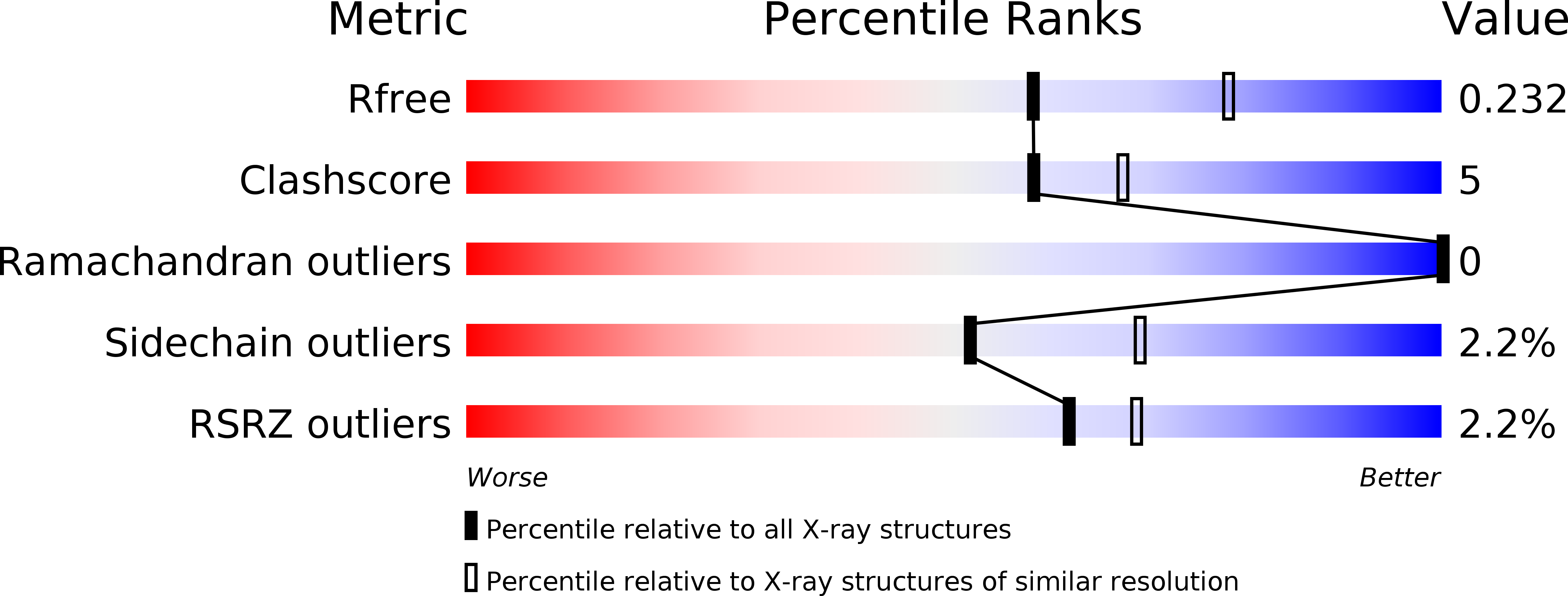
Experimental Data Snapshot
Entity ID: 1 | |||||
|---|---|---|---|---|---|
| Molecule | Chains | Sequence Length | Organism | Details | Image |
| Ac-ChiA | 551 | Autographa californica nucleopolyhedrovirus | Mutation(s): 0 Gene Names: ChiA, Ac-ChiA EC: 3.2.1.14 | 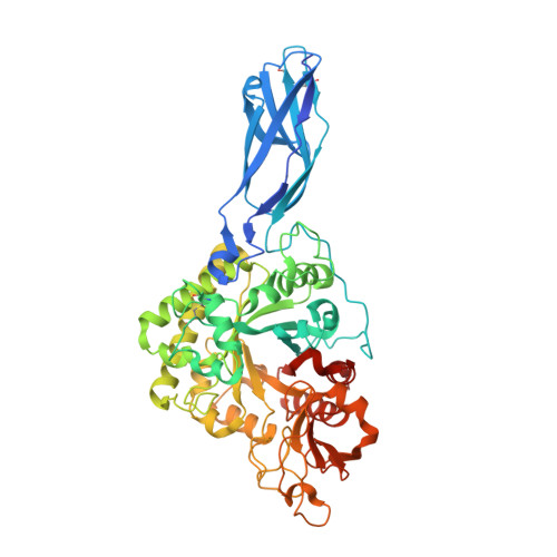 | |
UniProt | |||||
Find proteins for Q70SQ1 (Autographa californica nuclear polyhedrosis virus) Explore Q70SQ1 Go to UniProtKB: Q70SQ1 | |||||
Entity Groups | |||||
| Sequence Clusters | 30% Identity50% Identity70% Identity90% Identity95% Identity100% Identity | ||||
| UniProt Group | Q70SQ1 | ||||
Sequence AnnotationsExpand | |||||
| |||||
| Ligands 3 Unique | |||||
|---|---|---|---|---|---|
| ID | Chains | Name / Formula / InChI Key | 2D Diagram | 3D Interactions | |
| NAG Query on NAG | E [auth A] F [auth A] G [auth A] J [auth B] K [auth B] | 2-acetamido-2-deoxy-beta-D-glucopyranose C8 H15 N O6 OVRNDRQMDRJTHS-FMDGEEDCSA-N |  | ||
| SO4 Query on SO4 | N [auth B] | SULFATE ION O4 S QAOWNCQODCNURD-UHFFFAOYSA-L |  | ||
| CL Query on CL | H [auth A], I [auth A], O [auth B] | CHLORIDE ION Cl VEXZGXHMUGYJMC-UHFFFAOYSA-M |  | ||
| Length ( Å ) | Angle ( ˚ ) |
|---|---|
| a = 96.931 | α = 90 |
| b = 111.927 | β = 90 |
| c = 129.516 | γ = 90 |
| Software Name | Purpose |
|---|---|
| PHENIX | refinement |
| HKL-2000 | data reduction |
| HKL-2000 | data scaling |
| PHENIX | phasing |
| Funding Organization | Location | Grant Number |
|---|---|---|
| National Institutes of Health/National Institute of General Medical Sciences (NIH/NIGMS) | United States | P20GM103546 |
RCSB PDB (citation) is hosted by
RCSB PDB is a member of the


