5D68

Crystal structure of KRIT1 ARD-FERM

  • Classification: SIGNALING PROTEIN
  • Organism(s): Homo sapiens
  • Expression System: Escherichia coli
  • Mutation(s): No 

  • Deposited: 2015-08-11 Released: 2015-10-21 
  • Deposition Author(s): Zhang, R., Li, X., Boggon, T.J.
  • Funding Organization(s): National Institutes of Health/National Institute of Neurological Disorders and Stroke (NIH/NINDS), National Institutes of Health/National Institute of General Medical Sciences (NIH/NIGMS)

Experimental Data Snapshot

  • Method: X-RAY DIFFRACTION
  • Resolution: 2.91 Å
  • R-Value Free: 0.246 
  • R-Value Work: 0.207 
  • R-Value Observed: 0.209 

wwPDB Validation   3D Report Full Report


This is version 1.5 of the entry. See complete history


Literature

Structural analysis of the KRIT1 ankyrin repeat and FERM domains reveals a conformationally stable ARD-FERM interface.

Zhang, R.Li, X.Boggon, T.J.

(2015) J Struct Biol 192: 449-456

  • DOI: https://doi.org/10.1016/j.jsb.2015.10.006
  • Primary Citation of Related Structures:  
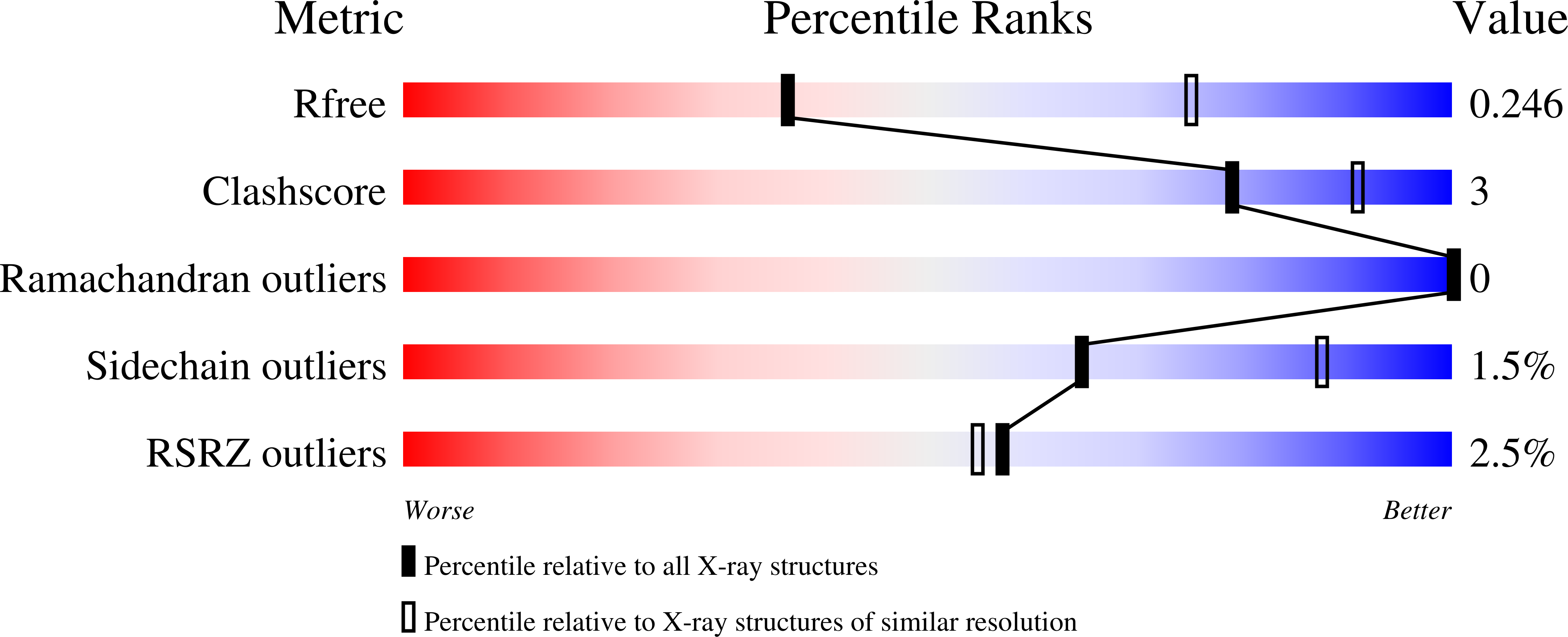
    5D68

  • PubMed Abstract: 

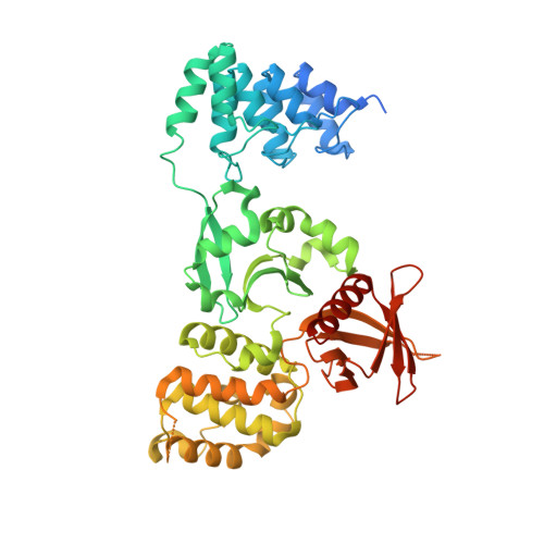
    Cerebral cavernous malformations (CCM) are vascular dysplasias that usually occur in the brain and are associated with mutations in the KRIT1/CCM1, CCM2/MGC4607/OSM/Malcavernin, and PDCD10/CCM3/TFAR15 genes. Here we report the 2.9 Å crystal structure of the ankyrin repeat domain (ARD) and FERM domain of the protein product of KRIT1 (KRIT1; Krev interaction trapped 1). The crystal structure reveals that the KRIT1 ARD contains 4 ankyrin repeats. There is an unusual conformation in the ANK4 repeat that is stabilized by Trp-404, and the structure reveals a solvent exposed ankyrin groove. Domain orientations of the three copies within the asymmetric unit suggest a stable interaction between KRIT1 ARD and FERM domains, indicating a globular ARD-FERM module. This resembles the additional F0 domain found N-terminal to the FERM domain of talin. Structural analysis of KRIT1 ARD-FERM highlights surface regions of high evolutionary conservation, and suggests potential sites that could mediate interaction with binding partners. The structure therefore provides a better understanding of KRIT1 at the molecular level.


  • Organizational Affiliation

    Department of Pharmacology, Yale University School of Medicine, 333 Cedar Street, New Haven, CT 06520, USA.


Macromolecules
Find similar proteins by:  (by identity cutoff)  |  3D Structure
Entity ID: 1
MoleculeChains Sequence LengthOrganismDetailsImage
Krev interaction trapped protein 1
A, B, C
486Homo sapiensMutation(s): 0 
Gene Names: KRIT1CCM1
UniProt & NIH Common Fund Data Resources
Find proteins for O00522 (Homo sapiens)
Explore O00522 
Go to UniProtKB:  O00522
PHAROS:  O00522
GTEx:  ENSG00000001631 
Entity Groups  
Sequence Clusters30% Identity50% Identity70% Identity90% Identity95% Identity100% Identity
UniProt GroupO00522
Sequence Annotations
Expand
  • Reference Sequence
Experimental Data & Validation

Experimental Data

Unit Cell:
Length ( Å )Angle ( ˚ )
a = 125.734α = 90
b = 80.242β = 91.69
c = 213.145γ = 90
Software Package:
Software NamePurpose
PHENIXrefinement
HKL-2000data reduction
HKL-2000data scaling
PHASERphasing

Structure Validation

View Full Validation Report


Entry History & Funding Information

Deposition Data


Funding OrganizationLocationGrant Number
National Institutes of Health/National Institute of Neurological Disorders and Stroke (NIH/NINDS)United StatesR01NS085078
National Institutes of Health/National Institute of General Medical Sciences (NIH/NIGMS)United StatesP41GM103403

Revision History  (Full details and data files)

  • Version 1.0: 2015-10-21
    Type: Initial release
  • Version 1.1: 2015-10-28
    Changes: Database references
  • Version 1.2: 2015-12-02
    Changes: Database references
  • Version 1.3: 2017-09-13
    Changes: Author supporting evidence, Database references, Derived calculations
  • Version 1.4: 2019-12-18
    Changes: Author supporting evidence
  • Version 1.5: 2023-09-27
    Changes: Data collection, Database references, Refinement description