5D62

MOA-Z-VAD-fmk complex, inverted orientation


Experimental Data Snapshot

  • Method: X-RAY DIFFRACTION
  • Resolution: 1.70 Å
  • R-Value Free: 0.198 
  • R-Value Work: 0.169 
  • R-Value Observed: 0.171 

wwPDB Validation   3D Report Full Report


Ligand Structure Quality Assessment 


This is version 2.1 of the entry. See complete history


Literature

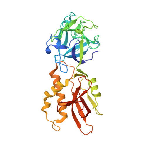
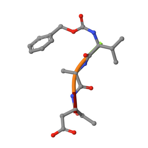
An Unusual Member of the Papain Superfamily: Mapping the Catalytic Cleft of the Marasmius oreades agglutinin (MOA) with a Caspase Inhibitor.

Cordara, G.van Eerde, A.Grahn, E.M.Winter, H.C.Goldstein, I.J.Krengel, U.

(2016) PLoS One 11: e0149407-e0149407

  • DOI: https://doi.org/10.1371/journal.pone.0149407
  • Primary Citation of Related Structures:  
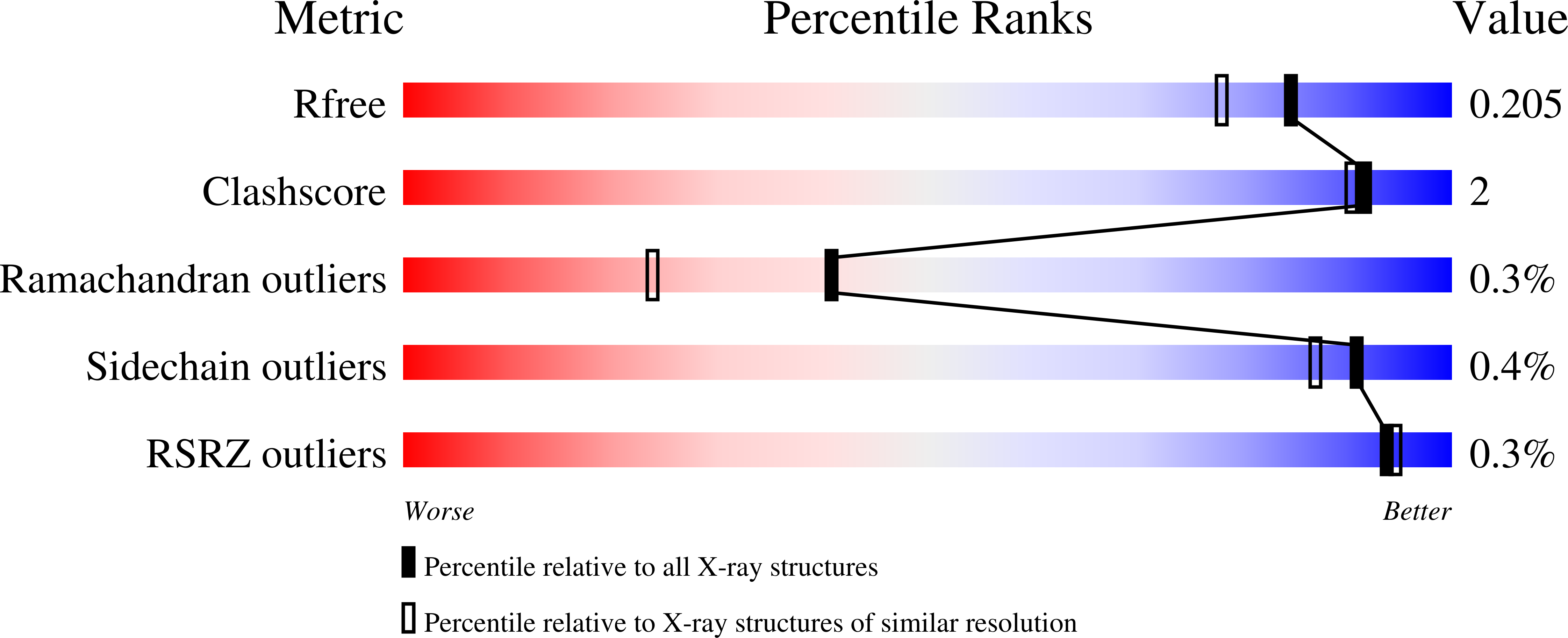
    5D61, 5D62, 5D63

  • PubMed Abstract: 

    Papain-like cysteine proteases (PLCPs) constitute the largest group of thiol-based protein degrading enzymes and are characterized by a highly conserved fold. They are found in bacteria, viruses, plants and animals and involved in a number of physiological and pathological processes, parasitic infections and host defense, making them interesting targets for drug design. The Marasmius oreades agglutinin (MOA) is a blood group B-specific fungal chimerolectin with calcium-dependent proteolytic activity. The proteolytic domain of MOA presents a unique structural arrangement, yet mimicking the main structural elements in known PLCPs. Here we present the X-ray crystal structure of MOA in complex with Z-VAD-fmk, an irreversible caspase inhibitor known to cross-react with PLCPs. The structural data allow modeling of the substrate binding geometry and mapping of the fundamental enzyme-substrate interactions. The new information consolidates MOA as a new, yet strongly atypical member of the papain superfamily. The reported complex is the first published structure of a PLCP in complex with the well characterized caspase inhibitor Z-VAD-fmk.


  • Organizational Affiliation

    Department of Chemistry, University of Oslo, Oslo, Norway.


Macromolecules
Find similar proteins by:  (by identity cutoff)  |  3D Structure
Entity ID: 1
MoleculeChains Sequence LengthOrganismDetailsImage
Agglutinin293Marasmius oreadesMutation(s): 0 
UniProt
Find proteins for Q8X123 (Marasmius oreades)
Explore Q8X123 
Go to UniProtKB:  Q8X123
Entity Groups  
Sequence Clusters30% Identity50% Identity70% Identity90% Identity95% Identity100% Identity
UniProt GroupQ8X123
Sequence Annotations
Expand
  • Reference Sequence

Find similar proteins by:  Sequence   |   3D Structure  

Entity ID: 2
MoleculeChains Sequence LengthOrganismDetailsImage
Z-VAD-fmkB [auth L]5synthetic constructMutation(s): 0 
Sequence Annotations
Expand
  • Reference Sequence
Oligosaccharides

Help

Entity ID: 3
MoleculeChains Length2D Diagram Glycosylation3D Interactions
alpha-L-fucopyranose-(1-2)-[alpha-D-galactopyranose-(1-3)]beta-D-galactopyranoseC [auth B]3N/A
Glycosylation Resources
GlyTouCan:  G47548GA
GlyCosmos:  G47548GA
GlyGen:  G47548GA
Entity ID: 4
MoleculeChains Length2D Diagram Glycosylation3D Interactions
alpha-D-galactopyranose-(1-3)-alpha-D-galactopyranoseD [auth C],
E [auth D]
2N/A
Glycosylation Resources
GlyTouCan:  G91563HD
GlyCosmos:  G91563HD
GlyGen:  G91563HD
Small Molecules
Ligands 5 Unique
IDChains Name / Formula / InChI Key2D Diagram3D Interactions
FUC
Query on FUC

Download Ideal Coordinates CCD File 
F [auth A],
G [auth A]
alpha-L-fucopyranose
C6 H12 O5
SHZGCJCMOBCMKK-SXUWKVJYSA-N
DMS
Query on DMS

Download Ideal Coordinates CCD File 
K [auth A],
L [auth A]
DIMETHYL SULFOXIDE
C2 H6 O S
IAZDPXIOMUYVGZ-UHFFFAOYSA-N
EDO
Query on EDO

Download Ideal Coordinates CCD File 
H [auth A],
I [auth A],
J [auth A]
1,2-ETHANEDIOL
C2 H6 O2
LYCAIKOWRPUZTN-UHFFFAOYSA-N
CA
Query on CA

Download Ideal Coordinates CCD File 
O [auth A],
P [auth A]
CALCIUM ION
Ca
BHPQYMZQTOCNFJ-UHFFFAOYSA-N
CL
Query on CL

Download Ideal Coordinates CCD File 
M [auth A],
N [auth A]
CHLORIDE ION
Cl
VEXZGXHMUGYJMC-UHFFFAOYSA-M
Experimental Data & Validation

Experimental Data

  • Method: X-RAY DIFFRACTION
  • Resolution: 1.70 Å
  • R-Value Free: 0.198 
  • R-Value Work: 0.169 
  • R-Value Observed: 0.171 
  • Space Group: P 63 2 2
Unit Cell:
Length ( Å )Angle ( ˚ )
a = 120.998α = 90
b = 120.998β = 90
c = 99.957γ = 120
Software Package:
Software NamePurpose
REFMACrefinement
XDSdata reduction
XSCALEdata scaling
PHASERphasing
Cootmodel building

Structure Validation

View Full Validation Report


Ligand Structure Quality Assessment 


Entry History 

Deposition Data

Revision History  (Full details and data files)

  • Version 1.0: 2016-03-02
    Type: Initial release
  • Version 1.1: 2016-12-21
    Changes: Non-polymer description
  • Version 2.0: 2020-07-29
    Type: Remediation
    Reason: Carbohydrate remediation
    Changes: Advisory, Atomic model, Data collection, Derived calculations, Structure summary
  • Version 2.1: 2024-01-10
    Changes: Data collection, Database references, Derived calculations, Refinement description, Structure summary