5CYV

Crystal structure of CouR from Rhodococcus jostii RHA1 bound to p-coumaroyl-CoA


Experimental Data Snapshot

  • Method: X-RAY DIFFRACTION
  • Resolution: 1.52 Å
  • R-Value Free: 0.202 
  • R-Value Work: 0.161 
  • R-Value Observed: 0.162 

wwPDB Validation   3D Report Full Report


Ligand Structure Quality Assessment 


This is version 1.6 of the entry. See complete history


Literature

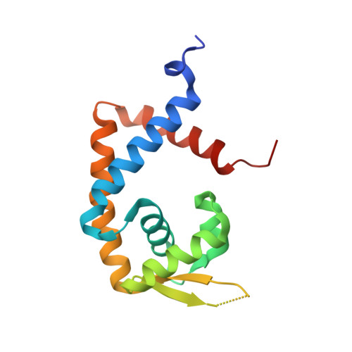
The activity of CouR, a MarR family transcriptional regulator, is modulated through a novel molecular mechanism.

Otani, H.Stogios, P.J.Xu, X.Nocek, B.Li, S.N.Savchenko, A.Eltis, L.D.

(2016) Nucleic Acids Res 44: 595-607

  • DOI: https://doi.org/10.1093/nar/gkv955
  • Primary Citation of Related Structures:  
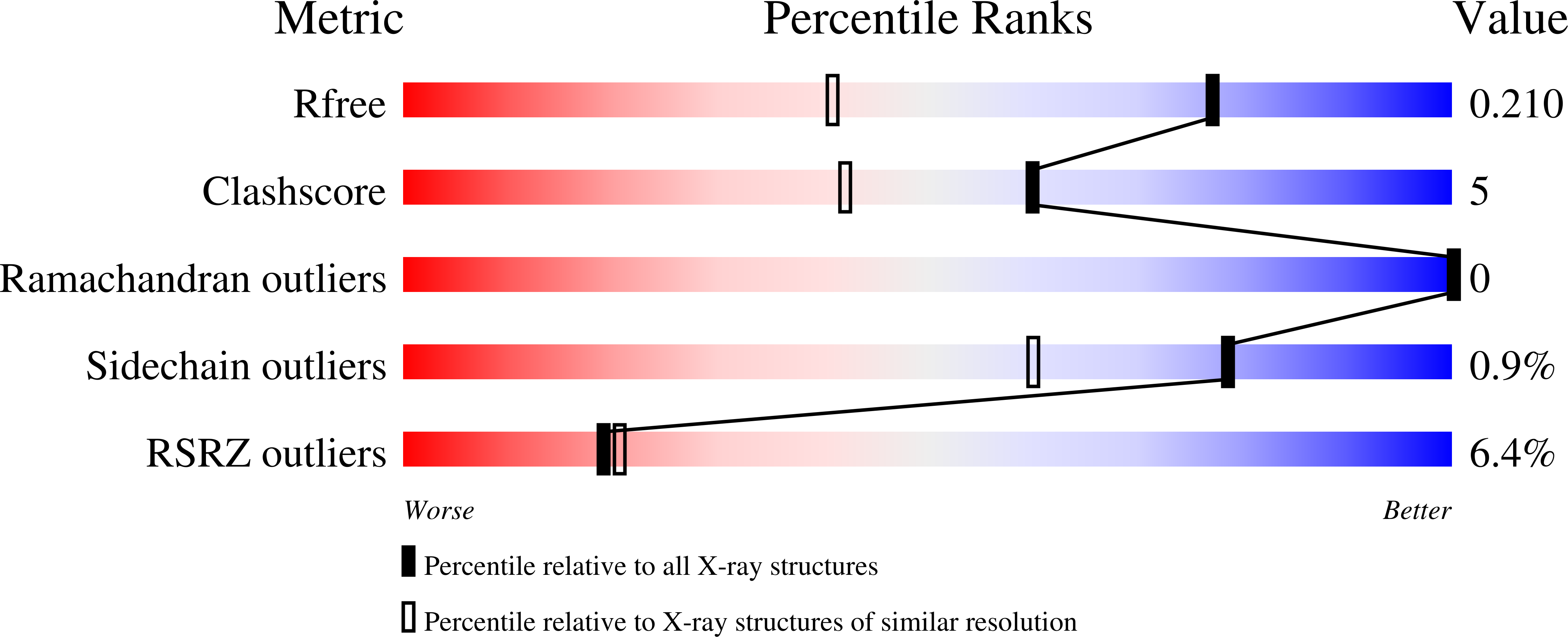
    5CYV

  • PubMed Abstract: 

    CouR, a MarR-type transcriptional repressor, regulates the cou genes, encoding p-hydroxycinnamate catabolism in the soil bacterium Rhodococcus jostii RHA1. The CouR dimer bound two molecules of the catabolite p-coumaroyl-CoA (Kd = 11 ± 1 μM). The presence of p-coumaroyl-CoA, but neither p-coumarate nor CoASH, abrogated CouR's binding to its operator DNA in vitro. The crystal structures of ligand-free CouR and its p-coumaroyl-CoA-bound form showed no significant conformational differences, in contrast to other MarR regulators. The CouR-p-coumaroyl-CoA structure revealed two ligand molecules bound to the CouR dimer with their phenolic moieties occupying equivalent hydrophobic pockets in each protomer and their CoA moieties adopting non-equivalent positions to mask the regulator's predicted DNA-binding surface. More specifically, the CoA phosphates formed salt bridges with predicted DNA-binding residues Arg36 and Arg38, changing the overall charge of the DNA-binding surface. The substitution of either arginine with alanine completely abrogated the ability of CouR to bind DNA. By contrast, the R36A/R38A double variant retained a relatively high affinity for p-coumaroyl-CoA (Kd = 89 ± 6 μM). Together, our data point to a novel mechanism of action in which the ligand abrogates the repressor's ability to bind DNA by steric occlusion of key DNA-binding residues and charge repulsion of the DNA backbone.


  • Organizational Affiliation

    Department of Microbiology and Immunology, Life Sciences Institute, The University of British Columbia, Vancouver, British Columbia V6T 1Z3, Canada.


Macromolecules
Find similar proteins by:  (by identity cutoff)  |  3D Structure
Entity ID: 1
MoleculeChains Sequence LengthOrganismDetailsImage
Transcriptional regulator
A, B
146Rhodococcus jostii RHA1Mutation(s): 0 
Gene Names: RHA1_ro05125
UniProt
Find proteins for Q0S6D0 (Rhodococcus jostii (strain RHA1))
Explore Q0S6D0 
Go to UniProtKB:  Q0S6D0
Entity Groups  
Sequence Clusters30% Identity50% Identity70% Identity90% Identity95% Identity100% Identity
UniProt GroupQ0S6D0
Sequence Annotations
Expand
  • Reference Sequence
Small Molecules
Ligands 4 Unique
IDChains Name / Formula / InChI Key2D Diagram3D Interactions
WCA
Query on WCA

Download Ideal Coordinates CCD File 
I [auth B],
J [auth B]
p-coumaroyl-CoA
C30 H42 N7 O18 P3 S
DMZOKBALNZWDKI-MATMFAIHSA-N
ACT
Query on ACT

Download Ideal Coordinates CCD File 
F [auth A]ACETATE ION
C2 H3 O2
QTBSBXVTEAMEQO-UHFFFAOYSA-M
CL
Query on CL

Download Ideal Coordinates CCD File 
G [auth A],
H [auth A],
K [auth B]
CHLORIDE ION
Cl
VEXZGXHMUGYJMC-UHFFFAOYSA-M
MG
Query on MG

Download Ideal Coordinates CCD File 
C [auth A],
D [auth A],
E [auth A]
MAGNESIUM ION
Mg
JLVVSXFLKOJNIY-UHFFFAOYSA-N
Binding Affinity Annotations 
IDSourceBinding Affinity
WCA Binding MOAD:  5CYV Kd: 1.10e+4 (nM) from 1 assay(s)
Experimental Data & Validation

Experimental Data

  • Method: X-RAY DIFFRACTION
  • Resolution: 1.52 Å
  • R-Value Free: 0.202 
  • R-Value Work: 0.161 
  • R-Value Observed: 0.162 
  • Space Group: C 2 2 21
Unit Cell:
Length ( Å )Angle ( ˚ )
a = 64.145α = 90
b = 134.385β = 90
c = 73.336γ = 90
Software Package:
Software NamePurpose
HKL-3000data reduction
HKL-3000data scaling
PHENIXphasing
PHENIXrefinement
Cootmodel building

Structure Validation

View Full Validation Report


Ligand Structure Quality Assessment 


Entry History & Funding Information

Deposition Data


Funding OrganizationLocationGrant Number
Department of Energy (DOE, United States)United StatesDE-AC02-06CH11357

Revision History  (Full details and data files)

  • Version 1.0: 2015-08-12
    Type: Initial release
  • Version 1.1: 2016-03-23
    Changes: Database references
  • Version 1.2: 2017-09-20
    Changes: Author supporting evidence, Database references, Derived calculations
  • Version 1.3: 2017-11-01
    Changes: Author supporting evidence
  • Version 1.4: 2019-12-04
    Changes: Author supporting evidence
  • Version 1.5: 2023-09-27
    Changes: Data collection, Database references, Derived calculations, Refinement description
  • Version 1.6: 2024-03-13
    Changes: Source and taxonomy