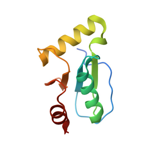
5CAX

CRYSTAL STRUCTURE OF METHANOSARCINA ACETIVORANS METHANOREDOXIN


Experimental Data Snapshot

  • Method: X-RAY DIFFRACTION
  • Resolution: 2.45 Å
  • R-Value Free: 0.272 
  • R-Value Work: 0.215 
  • R-Value Observed: 0.221 

wwPDB Validation   3D Report Full Report


This is version 1.3 of the entry. See complete history


Literature

Structural and Biochemical Characterizations of Methanoredoxin from Methanosarcina acetivorans, a Glutaredoxin-Like Enzyme with Coenzyme M-Dependent Protein Disulfide Reductase Activity.

Yenugudhati, D.Prakash, D.Kumar, A.K.Kumar, R.S.Yennawar, N.H.Yennawar, H.P.Ferry, J.G.

(2016) Biochemistry 55: 313-321

  • DOI: https://doi.org/10.1021/acs.biochem.5b00823
  • Primary Citation of Related Structures:  
    5CAX

  • PubMed Abstract: 

    Glutaredoxins (GRXs) are thiol-disulfide oxidoreductases abundant in prokaryotes, although little is understood of these enzymes from the domain Archaea. The numerous characterized GRXs from the domain Bacteria utilize a diversity of low-molecular-weight thiols in addition to glutathione as reductants. We report here the biochemical and structural properties of a GRX-like protein named methanoredoxin (MRX) from Methanosarcina acetivorans of the domain Archaea. MRX utilizes coenzyme M (CoMSH) as reductant for insulin disulfide reductase activity, which adds to the diversity of thiol protectants in prokaryotes. Cell-free extracts of M. acetivorans displayed CoMS-SCoM reductase activity that complements the CoMSH-dependent activity of MRX. The crystal structure exhibits a classic thioredoxin-glutaredoxin fold comprising three α-helices surrounding four antiparallel β-sheets. A pocket on the surface contains a CVWC motif, identifying the active site with architecture similar to GRXs. Although it is a monomer in solution, the crystal lattice has four monomers in a dimer of dimers arrangement. A cadmium ion is found within the active site of each monomer. Two such ions stabilize the N-terminal tails and dimer interfaces. Our modeling studies indicate that CoMSH and glutathione (GSH) bind to the active site of MRX similar to the binding of GSH in GRXs, although there are differences in the amino acid composition of the binding motifs. The results, combined with our bioinformatic analyses, show that MRX represents a class of GRX-like enzymes present in a diversity of methane-producing Archaea.


  • Organizational Affiliation

    Department of Biochemistry and Molecular Biology, ‡Huck Institutes of Life Sciences, The Pennsylvania State University , University Park, Pennsylvania 16802, United States.


Macromolecules
Find similar proteins by:  (by identity cutoff)  |  3D Structure
Entity ID: 1
MoleculeChains Sequence LengthOrganismDetailsImage
Glutaredoxin
A, B, C, D
101Methanosarcina acetivorans C2AMutation(s): 0 
Gene Names: MA_1658
UniProt
Find proteins for Q8TQ93 (Methanosarcina acetivorans (strain ATCC 35395 / DSM 2834 / JCM 12185 / C2A))
Explore Q8TQ93 
Go to UniProtKB:  Q8TQ93
Entity Groups  
Sequence Clusters30% Identity50% Identity70% Identity90% Identity95% Identity100% Identity
UniProt GroupQ8TQ93
Sequence Annotations
Expand
  • Reference Sequence
Small Molecules
Ligands 3 Unique
IDChains Name / Formula / InChI Key2D Diagram3D Interactions
CD
Query on CD

Download Ideal Coordinates CCD File 
E [auth A]
F [auth A]
G [auth A]
I [auth B]
J [auth B]
E [auth A],
F [auth A],
G [auth A],
I [auth B],
J [auth B],
K [auth B],
M [auth C],
N [auth C],
O [auth C],
P [auth D],
Q [auth D],
R [auth D]
CADMIUM ION
Cd
WLZRMCYVCSSEQC-UHFFFAOYSA-N
SO4
Query on SO4

Download Ideal Coordinates CCD File 
L [auth B],
V [auth D]
SULFATE ION
O4 S
QAOWNCQODCNURD-UHFFFAOYSA-L
ACY
Query on ACY

Download Ideal Coordinates CCD File 
H [auth A],
S [auth D],
T [auth D],
U [auth D]
ACETIC ACID
C2 H4 O2
QTBSBXVTEAMEQO-UHFFFAOYSA-N
Experimental Data & Validation

Experimental Data

  • Method: X-RAY DIFFRACTION
  • Resolution: 2.45 Å
  • R-Value Free: 0.272 
  • R-Value Work: 0.215 
  • R-Value Observed: 0.221 
  • Space Group: P 41
Unit Cell:
Length ( Å )Angle ( ˚ )
a = 59.154α = 90
b = 59.154β = 90
c = 172.978γ = 90
Software Package:
Software NamePurpose
PHENIXrefinement
HKL-2000data reduction
HKL-2000data scaling
PHENIXphasing

Structure Validation

View Full Validation Report


Entry History & Funding Information

Deposition Data


Funding OrganizationLocationGrant Number
Department of Energy (DOE, United States)United StatesDE-FG02-95ER20198 MOD16

Revision History  (Full details and data files)

  • Version 1.0: 2016-02-24
    Type: Initial release
  • Version 1.1: 2017-09-27
    Changes: Author supporting evidence, Derived calculations
  • Version 1.2: 2019-12-04
    Changes: Author supporting evidence
  • Version 1.3: 2023-09-27
    Changes: Data collection, Database references, Derived calculations, Refinement description