


Enhancing the Thermostability of beta-Glucuronidase by Rationally Redesigning the Catalytic Domain Based on Sequence Alignment Strategy
Feng, X.D., Tang, H., Han, B.J., Lv, B., Li, C.(2016) Ind Eng Chem Res 55: 5474-5483
Experimental Data Snapshot
wwPDB Validation 3D Report Full Report

(2016) Ind Eng Chem Res 55: 5474-5483
Entity ID: 1 | |||||
|---|---|---|---|---|---|
| Molecule | Chains | Sequence Length | Organism | Details | Image |
| Glucuronidase | 612 | Aspergillus oryzae | Mutation(s): 0 Gene Names: Pgus EC: 3.2.1.25 | 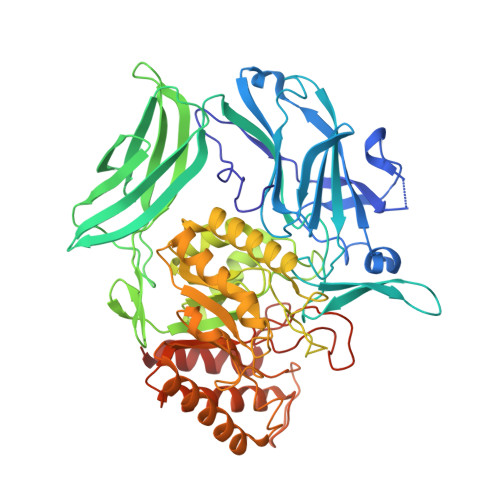 | |
UniProt | |||||
Find proteins for A7XS03 (Aspergillus oryzae) Explore A7XS03 Go to UniProtKB: A7XS03 | |||||
Entity Groups | |||||
| Sequence Clusters | 30% Identity50% Identity70% Identity90% Identity95% Identity100% Identity | ||||
| UniProt Group | A7XS03 | ||||
Sequence AnnotationsExpand | |||||
| |||||
| Length ( Å ) | Angle ( ˚ ) |
|---|---|
| a = 110.32 | α = 90 |
| b = 110.32 | β = 90 |
| c = 480.722 | γ = 120 |
| Software Name | Purpose |
|---|---|
| HKL-2000 | data processing |
| HKL-2000 | data scaling |
| AMoRE | phasing |
| REFMAC | refinement |
| Coot | model building |
RCSB PDB (citation) is hosted by
RCSB PDB is a member of the

