5BND

Crystal structure of the C-terminal domain of TagH


Experimental Data Snapshot

  • Method: X-RAY DIFFRACTION
  • Resolution: 2.68 Å
  • R-Value Free: 0.257 
  • R-Value Work: 0.206 
  • R-Value Observed: 0.208 

wwPDB Validation   3D Report Full Report


This is version 1.2 of the entry. See complete history


Literature

SH3-Like Motif-Containing C-terminal Domain of Staphylococcal Teichoic Acid Transporter Suggests Possible Function.

Ko, T.P.Tseng, S.T.Lai, S.J.Chen, S.C.Guan, H.H.Yang, C.S.Chen, C.J.Chen, Y.

(2016) Proteins 

  • DOI: https://doi.org/10.1002/prot.25074
  • Primary Citation of Related Structures:  
    5BND

  • PubMed Abstract: 

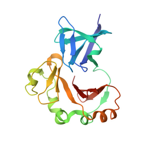
    The negatively charged bacterial polysaccharides-wall teichoic acids (WTAs) are synthesized intracellularly and exported by a two-component transporter, TagGH, comprising a transmembrane subunit TagG and an ATPase subunit TagH. We determined the crystal structure of the C-terminal domain of TagH (TagH-C) to investigate its function. The structure shows an N-terminal SH3-like subdomain wrapped by a C-terminal subdomain with an anti-parallel β-sheet and an outer shell of α-helices. A stretch of positively charged surface across the subdomain interface is flanked by two negatively charged regions, suggesting a potential binding site for negatively charged polymers, such as WTAs or acidic peptide chains. Proteins 2016; 84:1328-1332. © 2016 Wiley Periodicals, Inc.


  • Organizational Affiliation

    Institute of Biological Chemistry, Academia Sinica, Taipei, Taiwan.


Macromolecules
Find similar proteins by:  (by identity cutoff)  |  3D Structure
Entity ID: 1
MoleculeChains Sequence LengthOrganismDetailsImage
ABC transporter, ATP-binding protein
A, B, C
179Staphylococcus epidermidis RP62AMutation(s): 0 
Gene Names: SERP1409
UniProt
Find proteins for Q5HN63 (Staphylococcus epidermidis (strain ATCC 35984 / RP62A))
Explore Q5HN63 
Go to UniProtKB:  Q5HN63
Entity Groups  
Sequence Clusters30% Identity50% Identity70% Identity90% Identity95% Identity100% Identity
UniProt GroupQ5HN63
Sequence Annotations
Expand
  • Reference Sequence
Experimental Data & Validation

Experimental Data

  • Method: X-RAY DIFFRACTION
  • Resolution: 2.68 Å
  • R-Value Free: 0.257 
  • R-Value Work: 0.206 
  • R-Value Observed: 0.208 
  • Space Group: P 21 21 2
Unit Cell:
Length ( Å )Angle ( ˚ )
a = 104.576α = 90
b = 124.749β = 90
c = 56.132γ = 90
Software Package:
Software NamePurpose
PHENIXrefinement
HKL-2000data reduction
HKL-2000data scaling
PHENIXphasing

Structure Validation

View Full Validation Report


Entry History 

Deposition Data

Revision History  (Full details and data files)

  • Version 1.0: 2016-07-06
    Type: Initial release
  • Version 1.1: 2017-09-27
    Changes: Data collection, Database references, Derived calculations
  • Version 1.2: 2024-03-20
    Changes: Data collection, Database references