Structural and Functional Analyses of Periplasmic 5'-Methylthioadenosine/S-Adenosylhomocysteine Nucleosidase from Aeromonas hydrophila.
Xu, Y., Wang, L., Chen, J., Zhao, J., Fan, S., Dong, Y., Ha, N.C., Quan, C.(2017) Biochemistry 56: 5347-5355
- PubMed: 28862845
- DOI: https://doi.org/10.1021/acs.biochem.7b00691
- Primary Citation of Related Structures:
5B7G, 5B7N, 5B7P, 5B7Q - PubMed Abstract:
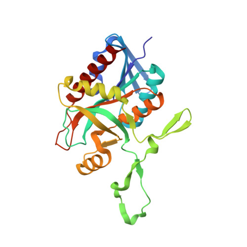
The Gram-negative, rod-shaped bacterium Aeromonas hydrophila has two multifunctional 5'-methylthioadenosine/S-adenosylhomocysteine nucleosidase (MTAN) enzymes, MtaN-1 and MtaN-2, that differ from those in other bacteria. These proteins are essential for several metabolic pathways, including biological methylation, polyamine biosynthesis, methionine recycling, and bacterial quorum sensing. To gain insight into how these two proteins function, we determined four high-resolution crystal structures of MtaN-1 in its apo form and in complex with the substrates S-adenosyl-l-homocysteine, 5'-methylthioadenosine, and 5'-deoxyadenosine. We found that the domain structures were generally similar, although slight differences were evident. The crystal structure demonstrates that AhMtaN-1 has an extension of the binding pocket and revealed that a tryptophan in the active site (Trp199) may play a major role in substrate binding, unlike in other MTAN proteins. Mutation of the Trp199 residue completely abolished the enzyme activity. Trp199 was identified as an active site residue that is essential for catalysis. Furthermore, biochemical characterization of AhMtaN-1 and AhMtaN-2 demonstrated that AhMtaN-1 exhibits inherent trypsin resistance that is higher than that of AhMtaN-2. Additionally, the thermally unfolded AhMtaN-2 protein is capable of refolding into active forms, whereas the thermally unfolded AhMtaN-1 protein does not have this ability. Examining the different biochemical characteristics related to the functional roles of AhMtaN-1 and AhMtaN-2 would be interesting. Indeed, the biochemical characterization of these structural features would provide a structural basis for the design of new antibiotics against A. hydrophila.
Organizational Affiliation:
Department of Bioengineering, College of Life Science, Dalian Minzu University , Dalian 116600, Liaoning, China.