5B6P

Structure of the dodecameric type-II dehydrogenate dehydratase from Acinetobacter baumannii at 2.00 A resolution


Experimental Data Snapshot

  • Method: X-RAY DIFFRACTION
  • Resolution: 2.00 Å
  • R-Value Free: 0.232 
  • R-Value Work: 0.201 
  • R-Value Observed: 0.201 

wwPDB Validation   3D Report Full Report


This is version 1.2 of the entry. See complete history


Literature

Binding studies and structure determination of the recombinantly produced type-II 3-dehydroquinate dehydratase from Acinetobacter baumannii.

Iqbal, N.Kumar, M.Sharma, P.Yadav, S.P.Kaur, P.Sharma, S.Singh, T.P.

(2017) Int J Biol Macromol 94: 459-465

  • DOI: https://doi.org/10.1016/j.ijbiomac.2016.10.049
  • Primary Citation of Related Structures:  
    5B6P

  • PubMed Abstract: 

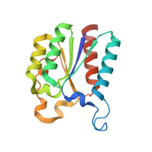
    Dehydroquinase (3-dehydroquinate dehydratase, DHQD, EC 4.2.1.10) catalyzes the conversion of dehydroquinate to dehydroshikimate. DHQD from Acinetobacter baumannii (AbDHQD) was cloned, expressed and purified to homogeneity. The binding studies showed that two compounds quinic acid and citrazinic acid bound to AbDHQD at micromolar concentrations. AbDHQD was crystallized using 30% PEG-3350, 50mM tris-HCl and 1.0M MgSO4 at pH 8.0. Crystals of AbDHQD were stabilized with 25% glycerol for data collection at 100K. The X-ray intensity data were collected to 2.0Å resolution. Crystals belonged to monoclinic space group P2 1 with cell dimensions, a=82.3, b=95.3, c=132.3Å and β=95.7°. The structure was solved with molecular replacement method and refined to values of 0.200 and 0.232 for R cryst and R free factors. The structures of 12 crystallographically independent molecules in the asymmetry unit were identical with r.m.s shifts for the C α atoms ranging from 0.3Å to 0.8Å. They formed a dodecamer with four trimers arranged in a tetrahedral manner. The classical lid adopted an open conformation although a sulfate ion was observed in the substrate binding site. As a result of which, the compounds quinic acid and citrazinic acid could not bind to AbDHQD.


  • Organizational Affiliation

    Department of Biophysics, All India Institute of Medical Sciences, New Delhi, India.


Macromolecules
Find similar proteins by:  (by identity cutoff)  |  3D Structure
Entity ID: 1
MoleculeChains Sequence LengthOrganismDetailsImage
3-dehydroquinate dehydratase
A, B, C, D, E
A, B, C, D, E, F, G, H, I, J, K, L
151Acinetobacter baumannii ATCC 17978Mutation(s): 0 
Gene Names: aroQA1S_2009
EC: 4.2.1.10
UniProt
Find proteins for A3M692 (Acinetobacter baumannii (strain ATCC 17978 / CIP 53.77 / LMG 1025 / NCDC KC755 / 5377))
Explore A3M692 
Go to UniProtKB:  A3M692
Entity Groups  
Sequence Clusters30% Identity50% Identity70% Identity90% Identity95% Identity100% Identity
UniProt GroupA3M692
Sequence Annotations
Expand
  • Reference Sequence
Small Molecules
Ligands 1 Unique
IDChains Name / Formula / InChI Key2D Diagram3D Interactions
SO4
Query on SO4

Download Ideal Coordinates CCD File 
M [auth A]
N [auth B]
O [auth C]
P [auth D]
Q [auth E]
M [auth A],
N [auth B],
O [auth C],
P [auth D],
Q [auth E],
R [auth F],
S [auth G],
T [auth H],
U [auth I],
V [auth J],
W [auth K],
X [auth L]
SULFATE ION
O4 S
QAOWNCQODCNURD-UHFFFAOYSA-L
Experimental Data & Validation

Experimental Data

  • Method: X-RAY DIFFRACTION
  • Resolution: 2.00 Å
  • R-Value Free: 0.232 
  • R-Value Work: 0.201 
  • R-Value Observed: 0.201 
  • Space Group: P 1 21 1
Unit Cell:
Length ( Å )Angle ( ˚ )
a = 82.284α = 90
b = 95.279β = 95.73
c = 132.263γ = 90
Software Package:
Software NamePurpose
REFMACrefinement
HKL-2000data reduction
HKL-2000data scaling
MOLREPphasing

Structure Validation

View Full Validation Report


Entry History 

Deposition Data

Revision History  (Full details and data files)

  • Version 1.0: 2016-09-07
    Type: Initial release
  • Version 1.1: 2019-04-17
    Changes: Data collection, Database references, Derived calculations
  • Version 1.2: 2023-11-08
    Changes: Data collection, Database references, Refinement description