


Structure and function of a prenyltransferase
Zhang, L., Chen, C.-C., Ko, T.-P., Guo, R.-T., Oldfield, E.O.To be published.
Experimental Data Snapshot
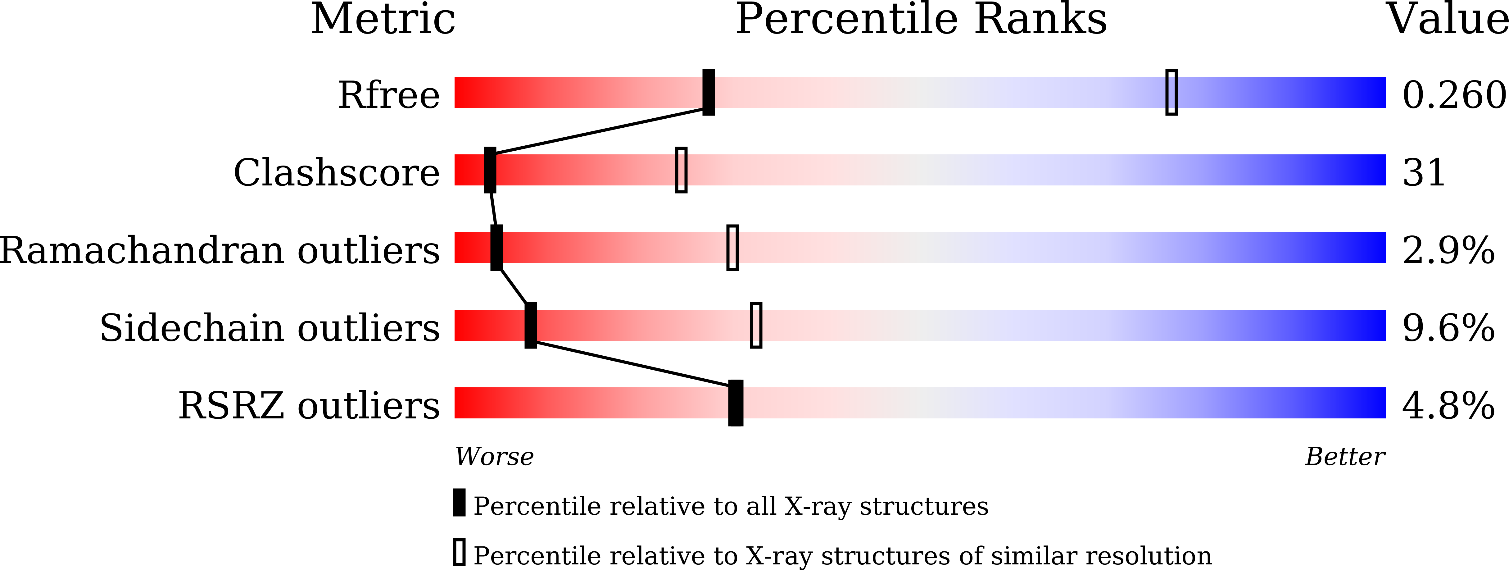
wwPDB Validation 3D Report Full Report
Entity ID: 1 | |||||
|---|---|---|---|---|---|
| Molecule | Chains | Sequence Length | Organism | Details | Image |
| MoeN5 | 294 | Streptomyces viridosporus | Mutation(s): 0 Gene Names: moeN5 | 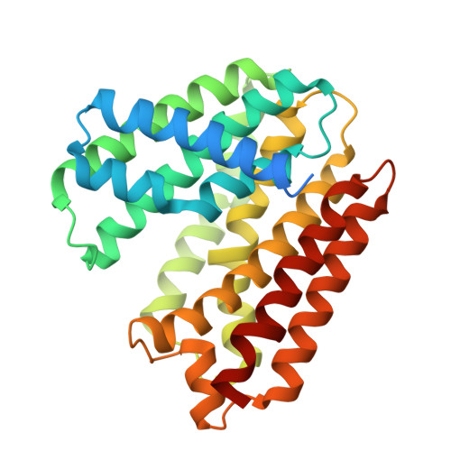 | |
UniProt | |||||
Find proteins for A0A010 (Streptomyces viridosporus) Explore A0A010 Go to UniProtKB: A0A010 | |||||
Entity Groups | |||||
| Sequence Clusters | 30% Identity50% Identity70% Identity90% Identity95% Identity100% Identity | ||||
| UniProt Group | A0A010 | ||||
Sequence AnnotationsExpand | |||||
| |||||
| Length ( Å ) | Angle ( ˚ ) |
|---|---|
| a = 106.604 | α = 90 |
| b = 106.604 | β = 90 |
| c = 310.713 | γ = 90 |
| Software Name | Purpose |
|---|---|
| CNS | refinement |
| HKL-2000 | data reduction |
| HKL-2000 | data scaling |
| CNS | phasing |
RCSB PDB (citation) is hosted by
RCSB PDB is a member of the

