5AOV

Ternary Crystal Structure of Pyrococcus furiosus Glyoxylate Hydroxypyruvate Reductase in presence of glyoxylate


Experimental Data Snapshot

  • Method: X-RAY DIFFRACTION
  • Resolution: 1.40 Å
  • R-Value Free: 0.143 
  • R-Value Work: 0.131 
  • R-Value Observed: 0.132 

wwPDB Validation   3D Report Full Report


Ligand Structure Quality Assessment 


This is version 2.0 of the entry. See complete history


Literature

New Insights Into the Mechanism of Substrates Trafficking in Glyoxylate/Hydroxypyruvate Reductases.

Lassalle, L.Engilberge, S.Madern, D.Vauclare, P.Franzetti, B.Girard, E.

(2016) Sci Rep 6: 20629

  • DOI: https://doi.org/10.1038/srep20629
  • Primary Citation of Related Structures:  
    5AOV, 6BII

  • PubMed Abstract: 

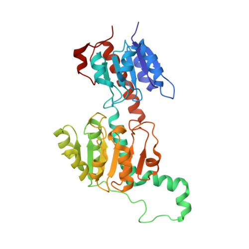
    Glyoxylate accumulation within cells is highly toxic. In humans, it is associated with hyperoxaluria type 2 (PH2) leading to renal failure. The glyoxylate content within cells is regulated by the NADPH/NADH dependent glyoxylate/hydroxypyruvate reductases (GRHPR). These are highly conserved enzymes with a dual activity as they are able to reduce glyoxylate to glycolate and to convert hydroxypyruvate into D-glycerate. Despite the determination of high-resolution X-ray structures, the substrate recognition mode of this class of enzymes remains unclear. We determined the structure at 2.0 Å resolution of a thermostable GRHPR from Archaea as a ternary complex in the presence of D-glycerate and NADPH. This shows a binding mode conserved between human and archeal enzymes. We also determined the first structure of GRHPR in presence of glyoxylate at 1.40 Å resolution. This revealed the pivotal role of Leu53 and Trp138 in substrate trafficking. These residues act as gatekeepers at the entrance of a tunnel connecting the active site to protein surface. Taken together, these results allowed us to propose a general model for GRHPR mode of action.


  • Organizational Affiliation

    Univ. Grenoble Alpes, IBS, F-38044 Grenoble, France.


Macromolecules
Find similar proteins by:  (by identity cutoff)  |  3D Structure
Entity ID: 1
MoleculeChains Sequence LengthOrganismDetailsImage
GLYOXYLATE REDUCTASE336Pyrococcus furiosusMutation(s): 0 
EC: 1.1.1.26 (PDB Primary Data), 1.1.1.81 (PDB Primary Data), 1.1.1.79 (PDB Primary Data)
UniProt
Find proteins for Q8U3Y2 (Pyrococcus furiosus (strain ATCC 43587 / DSM 3638 / JCM 8422 / Vc1))
Explore Q8U3Y2 
Go to UniProtKB:  Q8U3Y2
Entity Groups  
Sequence Clusters30% Identity50% Identity70% Identity90% Identity95% Identity100% Identity
UniProt GroupQ8U3Y2
Sequence Annotations
Expand
  • Reference Sequence
Small Molecules
Ligands 5 Unique
IDChains Name / Formula / InChI Key2D Diagram3D Interactions
NAP
Query on NAP

Download Ideal Coordinates CCD File 
B [auth A]NADP NICOTINAMIDE-ADENINE-DINUCLEOTIDE PHOSPHATE
C21 H28 N7 O17 P3
XJLXINKUBYWONI-NNYOXOHSSA-N
1PE
Query on 1PE

Download Ideal Coordinates CCD File 
D [auth A]
E [auth A]
F [auth A]
G [auth A]
H [auth A]
D [auth A],
E [auth A],
F [auth A],
G [auth A],
H [auth A],
I [auth A],
J [auth A],
K [auth A],
L [auth A],
M [auth A],
N [auth A],
O [auth A],
P [auth A],
V [auth A],
Z [auth A]
PENTAETHYLENE GLYCOL
C10 H22 O6
JLFNLZLINWHATN-UHFFFAOYSA-N
GLV
Query on GLV

Download Ideal Coordinates CCD File 
C [auth A],
W [auth A]
GLYOXYLIC ACID
C2 H2 O3
HHLFWLYXYJOTON-UHFFFAOYSA-N
EDO
Query on EDO

Download Ideal Coordinates CCD File 
Q [auth A],
R [auth A],
S [auth A],
T [auth A],
U [auth A]
1,2-ETHANEDIOL
C2 H6 O2
LYCAIKOWRPUZTN-UHFFFAOYSA-N
ACY
Query on ACY

Download Ideal Coordinates CCD File 
X [auth A],
Y [auth A]
ACETIC ACID
C2 H4 O2
QTBSBXVTEAMEQO-UHFFFAOYSA-N
Experimental Data & Validation

Experimental Data

  • Method: X-RAY DIFFRACTION
  • Resolution: 1.40 Å
  • R-Value Free: 0.143 
  • R-Value Work: 0.131 
  • R-Value Observed: 0.132 
  • Space Group: I 41
Unit Cell:
Length ( Å )Angle ( ˚ )
a = 114.579α = 90
b = 114.579β = 90
c = 118.123γ = 90
Software Package:
Software NamePurpose
PHENIXrefinement
XDSdata reduction
SCALAdata scaling
SHELXphasing

Structure Validation

View Full Validation Report


Ligand Structure Quality Assessment 


Entry History 

Deposition Data

Revision History  (Full details and data files)

  • Version 1.0: 2016-03-02
    Type: Initial release
  • Version 2.0: 2019-10-23
    Changes: Atomic model, Data collection, Other