5AN7

Structure of the engineered retro-aldolase RA95.5-8F with a bound 1,3-diketone inhibitor


Experimental Data Snapshot

  • Method: X-RAY DIFFRACTION
  • Resolution: 1.10 Å
  • R-Value Free: 0.188 
  • R-Value Observed: 0.136 

wwPDB Validation   3D Report Full Report


Ligand Structure Quality Assessment 


This is version 2.1 of the entry. See complete history


Literature

Emergence of a catalytic tetrad during evolution of a highly active artificial aldolase.

Obexer, R.Godina, A.Garrabou, X.Mittl, P.R.Baker, D.Griffiths, A.D.Hilvert, D.

(2017) Nat Chem 9: 50-56

  • DOI: https://doi.org/10.1038/nchem.2596
  • Primary Citation of Related Structures:  
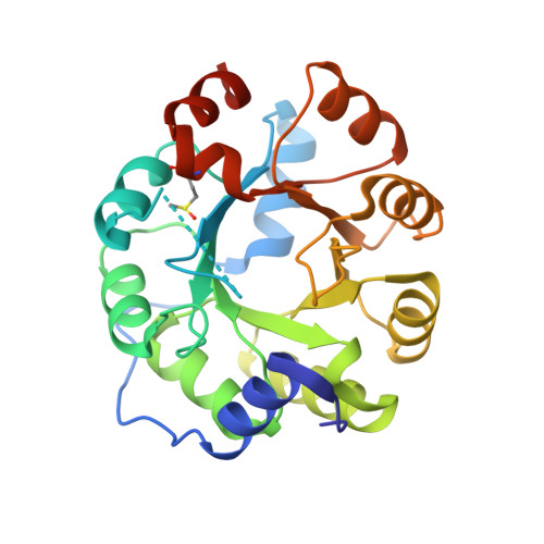
    5AN7, 5AOU

  • PubMed Abstract: 

    Designing catalysts that achieve the rates and selectivities of natural enzymes is a long-standing goal in protein chemistry. Here, we show that an ultrahigh-throughput droplet-based microfluidic screening platform can be used to improve a previously optimized artificial aldolase by an additional factor of 30 to give a >10 9 rate enhancement that rivals the efficiency of class I aldolases. The resulting enzyme catalyses a reversible aldol reaction with high stereoselectivity and tolerates a broad range of substrates. Biochemical and structural studies show that catalysis depends on a Lys-Tyr-Asn-Tyr tetrad that emerged adjacent to a computationally designed hydrophobic pocket during directed evolution. This constellation of residues is poised to activate the substrate by Schiff base formation, promote mechanistically important proton transfers and stabilize multiple transition states along a complex reaction coordinate. The emergence of such a sophisticated catalytic centre shows that there is nothing magical about the catalytic activities or mechanisms of naturally occurring enzymes, or the evolutionary process that gave rise to them.


  • Organizational Affiliation

    Laboratory of Organic Chemistry, ETH Zürich, 8093 Zürich, Switzerland.


Macromolecules
Find similar proteins by:  (by identity cutoff)  |  3D Structure
Entity ID: 1
MoleculeChains Sequence LengthOrganismDetailsImage
RA95.5-8F258Saccharolobus solfataricusMutation(s): 0 
UniProt
Find proteins for Q06121 (Saccharolobus solfataricus (strain ATCC 35092 / DSM 1617 / JCM 11322 / P2))
Explore Q06121 
Go to UniProtKB:  Q06121
Entity Groups  
Sequence Clusters30% Identity50% Identity70% Identity90% Identity95% Identity100% Identity
UniProt GroupQ06121
Sequence Annotations
Expand
  • Reference Sequence
Small Molecules
Modified Residues  1 Unique
IDChains TypeFormula2D DiagramParent
MHO
Query on MHO
A
L-PEPTIDE LINKINGC5 H11 N O3 SMET
Experimental Data & Validation

Experimental Data

  • Method: X-RAY DIFFRACTION
  • Resolution: 1.10 Å
  • R-Value Free: 0.188 
  • R-Value Observed: 0.136 
  • Space Group: P 21 21 2
Unit Cell:
Length ( Å )Angle ( ˚ )
a = 75.11α = 90
b = 84.43β = 90
c = 37.64γ = 90
Software Package:
Software NamePurpose
SHELXLrefinement
PHASERphasing

Structure Validation

View Full Validation Report


Ligand Structure Quality Assessment 


Entry History 

Deposition Data

Revision History  (Full details and data files)

  • Version 1.0: 2016-08-17
    Type: Initial release
  • Version 1.1: 2017-03-01
    Changes: Database references
  • Version 1.2: 2019-05-08
    Changes: Data collection, Derived calculations, Experimental preparation
  • Version 1.3: 2019-05-22
    Changes: Data collection, Refinement description
  • Version 2.0: 2019-10-23
    Changes: Atomic model, Data collection, Other
  • Version 2.1: 2024-01-10
    Changes: Data collection, Database references, Derived calculations, Refinement description