


Crystallographic Trapping of a Covalently Modified Heme in a Dye-Decolorizing Peroxidase
Strittmatter, E., Piontek, K., Plattner, D.A.To be published.
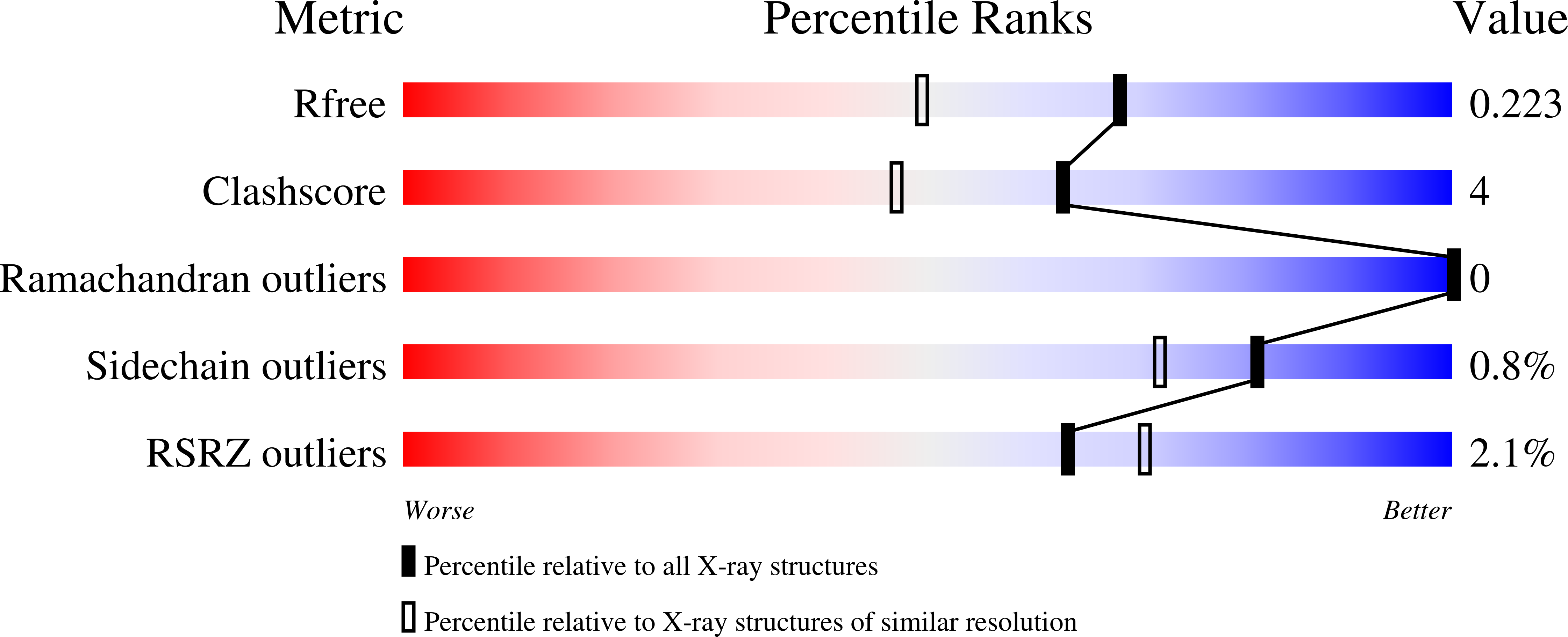
Experimental Data Snapshot
Entity ID: 1 | |||||
|---|---|---|---|---|---|
| Molecule | Chains | Sequence Length | Organism | Details | Image |
| DYE-DECOLORIZING PEROXIDASE | 446 | Auricularia auricula-judae | Mutation(s): 0 EC: 1.11.1.19 | 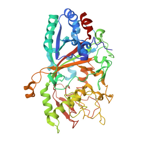 | |
UniProt | |||||
Find proteins for I2DBY1 (Auricularia auricula-judae) Explore I2DBY1 Go to UniProtKB: I2DBY1 | |||||
Entity Groups | |||||
| Sequence Clusters | 30% Identity50% Identity70% Identity90% Identity95% Identity100% Identity | ||||
| UniProt Group | I2DBY1 | ||||
Sequence AnnotationsExpand | |||||
| |||||
| Ligands 6 Unique | |||||
|---|---|---|---|---|---|
| ID | Chains | Name / Formula / InChI Key | 2D Diagram | 3D Interactions | |
| HEM Query on HEM | D [auth A], T [auth B] | PROTOPORPHYRIN IX CONTAINING FE C34 H32 Fe N4 O4 KABFMIBPWCXCRK-RGGAHWMASA-L |  | ||
| NAG Query on NAG | E [auth A], F [auth A], U [auth B], V [auth B] | 2-acetamido-2-deoxy-beta-D-glucopyranose C8 H15 N O6 OVRNDRQMDRJTHS-FMDGEEDCSA-N |  | ||
| CAC Query on CAC | G [auth A], W [auth B] | CACODYLATE ION C2 H6 As O2 OGGXGZAMXPVRFZ-UHFFFAOYSA-M |  | ||
| GOL Query on GOL | AA [auth B] BA [auth B] CA [auth B] DA [auth B] J [auth A] | GLYCEROL C3 H8 O3 PEDCQBHIVMGVHV-UHFFFAOYSA-N |  | ||
| GOA Query on GOA | K [auth A] | GLYCOLIC ACID C2 H4 O3 AEMRFAOFKBGASW-UHFFFAOYSA-N |  | ||
| FMT Query on FMT | EA [auth B], H [auth A], I [auth A], X [auth B] | FORMIC ACID C H2 O2 BDAGIHXWWSANSR-UHFFFAOYSA-N |  | ||
| Length ( Å ) | Angle ( ˚ ) |
|---|---|
| a = 65.84 | α = 90 |
| b = 46.57 | β = 100.49 |
| c = 147.63 | γ = 90 |
| Software Name | Purpose |
|---|---|
| REFMAC | refinement |
| XDS | data reduction |
| XDS | data scaling |
| PHASER | phasing |
RCSB PDB (citation) is hosted by
RCSB PDB is a member of the


