5ABV

Complex of D. melanogaster eIF4E with the 4E-binding protein Mextli


Experimental Data Snapshot

  • Method: X-RAY DIFFRACTION
  • Resolution: 2.13 Å
  • R-Value Free: 0.232 
  • R-Value Work: 0.192 
  • R-Value Observed: 0.194 

wwPDB Validation   3D Report Full Report


This is version 1.2 of the entry. See complete history


Literature

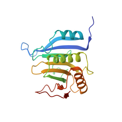
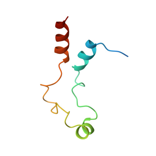
Mextli Proteins Use Both Canonical Bipartite and Novel Tripartite Binding Modes to Form Eif4E Complexes that Display Differential Sensitivity to 4E-BP Regulation

Peter, D.Weber, R.Koene, C.Chung, M.-Y.Ebertsch, L.Truffault, V.Weichenrieder, O.Igreja, C.Izaurralde, E.

(2015) Genes Dev 29: 1835

  • DOI: https://doi.org/10.1101/gad.269068.115
  • Primary Citation of Related Structures:  
    5ABU, 5ABV, 5ABX, 5ABY

  • PubMed Abstract: 

    The eIF4E-binding proteins (4E-BPs) are a diverse class of translation regulators that share a canonical eIF4E-binding motif (4E-BM) with eIF4G. Consequently, they compete with eIF4G for binding to eIF4E, thereby inhibiting translation initiation. Mextli (Mxt) is an unusual 4E-BP that promotes translation by also interacting with eIF3. Here we present the crystal structures of the eIF4E-binding regions of the Drosophila melanogaster (Dm) and Caenorhabditis elegans (Ce) Mxt proteins in complex with eIF4E in the cap-bound and cap-free states. The structures reveal unexpected evolutionary plasticity in the eIF4E-binding mode, with a classical bipartite interface for Ce Mxt and a novel tripartite interface for Dm Mxt. Both interfaces comprise a canonical helix and a noncanonical helix that engage the dorsal and lateral surfaces of eIF4E, respectively. Remarkably, Dm Mxt contains a C-terminal auxiliary helix that lies anti-parallel to the canonical helix on the eIF4E dorsal surface. In contrast to the eIF4G and Ce Mxt complexes, the Dm eIF4E-Mxt complexes are resistant to competition by bipartite 4E-BPs, suggesting that Dm Mxt can bind eIF4E when eIF4G binding is inhibited. Our results uncovered unexpected diversity in the binding modes of 4E-BPs, resulting in eIF4E complexes that display differential sensitivity to 4E-BP regulation.


  • Organizational Affiliation

    Department of Biochemistry, Max Planck Institute for Developmental Biology, 72076 Tübingen, Germany.


Macromolecules
Find similar proteins by:  (by identity cutoff)  |  3D Structure
Entity ID: 1
MoleculeChains Sequence LengthOrganismDetailsImage
EUKARYOTIC TRANSLATION INITIATION FACTOR 4E
A, C, E, G
184Drosophila melanogasterMutation(s): 0 
UniProt
Find proteins for P48598 (Drosophila melanogaster)
Explore P48598 
Go to UniProtKB:  P48598
Entity Groups  
Sequence Clusters30% Identity50% Identity70% Identity90% Identity95% Identity100% Identity
UniProt GroupP48598
Sequence Annotations
Expand
  • Reference Sequence
Find similar proteins by:  (by identity cutoff)  |  3D Structure
Entity ID: 2
MoleculeChains Sequence LengthOrganismDetailsImage
GH11071P
B, D, F, H
70Drosophila melanogasterMutation(s): 0 
UniProt
Find proteins for Q9VR35 (Drosophila melanogaster)
Explore Q9VR35 
Go to UniProtKB:  Q9VR35
Entity Groups  
Sequence Clusters30% Identity50% Identity70% Identity90% Identity95% Identity100% Identity
UniProt GroupQ9VR35
Sequence Annotations
Expand
  • Reference Sequence
Experimental Data & Validation

Experimental Data

  • Method: X-RAY DIFFRACTION
  • Resolution: 2.13 Å
  • R-Value Free: 0.232 
  • R-Value Work: 0.192 
  • R-Value Observed: 0.194 
  • Space Group: P 1 21 1
Unit Cell:
Length ( Å )Angle ( ˚ )
a = 65.9α = 90
b = 82.02β = 90.13
c = 84.73γ = 90
Software Package:
Software NamePurpose
BUSTERrefinement
XDSdata reduction
XSCALEdata scaling
PHASERphasing

Structure Validation

View Full Validation Report


Entry History 

Deposition Data

Revision History  (Full details and data files)

  • Version 1.0: 2015-09-02
    Type: Initial release
  • Version 1.1: 2015-09-23
    Changes: Database references
  • Version 1.2: 2024-01-10
    Changes: Data collection, Database references, Other, Refinement description