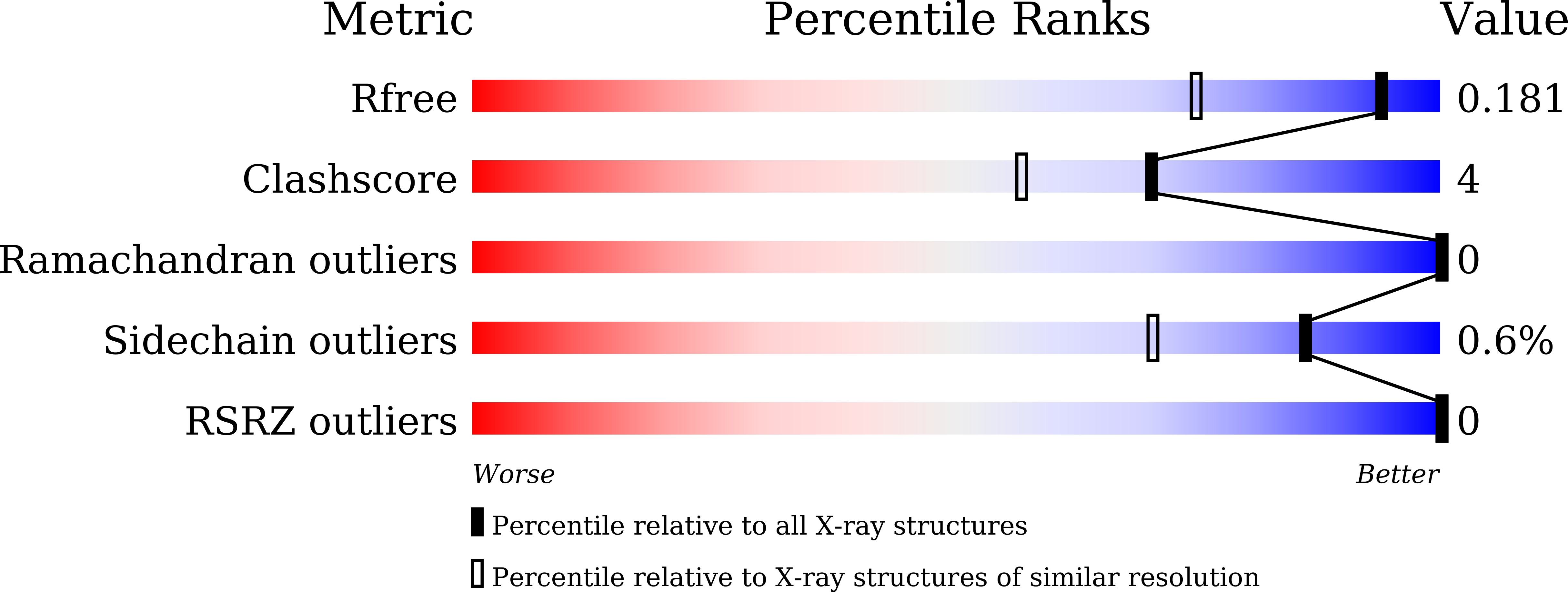
5ZWP

Crystal structure of the delta-class glutathione transferase from Musca domestica


Experimental Data Snapshot

  • Method: X-RAY DIFFRACTION
  • Resolution: 1.40 Å
  • R-Value Free: 0.176 
  • R-Value Work: 0.157 
  • R-Value Observed: 0.158 

wwPDB Validation   3D Report Full Report


Ligand Structure Quality Assessment 


This is version 1.2 of the entry. See complete history


Literature

Crystal structure of the delta-class glutathione transferase in Musca domestica

Sue, M.Yajima, S.

(2018) Biochem Biophys Res Commun 502: 345-350

  • DOI: https://doi.org/10.1016/j.bbrc.2018.05.161
  • Primary Citation of Related Structures:  
    5ZWP

  • PubMed Abstract: 

    Among the various glutathione transferase (GST) isozymes in insects, the delta- and epsilon-class GSTs fulfill critical functions during the detoxification of insecticides. We crystalized MdGSTD1, the major delta-class GST isozyme in the housefly (Musca domestica), in complex with glutathione (GSH) and solved its structure at a resolution of 1.4 Å. The overall folding of MdGSTD1 resembled other known delta-class GSTs. Its substrate binding pocket was exposed to solvent and considerably more open than in the epsilon-class GST from M. domestica (MdGSTE2). However, their C-terminal structures differed the most because of the different lengths of the C-terminal regions. Although this region does not seem to directly interact with substrates, its deletion reduced the enzymatic activity by more than 70%, indicating a function in maintaining the proper conformation of the binding pocket. Binding of GSH to the GSH-binding region of MdGSTD1 results in a rigid conformation of this region. Although MdGSTD1 has a higher affinity for GSH than the epsilon class enzymes, the thiol group of the GSH molecule was not close enough to serine residue 9 to form a hydrogen-bond with this residue, which is predicted to act as the catalytic center for thiol group deprotonation in GSH.


  • Organizational Affiliation

    Department of Agricultural Chemistry, Tokyo University of Agriculture, Sakuragaoka 1-1-1, Setagaya, Tokyo, 156-8502, Japan. Electronic address: sue@nodai.ac.jp.


Macromolecules
Find similar proteins by:  (by identity cutoff)  |  3D Structure
Entity ID: 1
MoleculeChains Sequence LengthOrganismDetailsImage
Glutathione S-transferase 1
A, B
208Musca domesticaMutation(s): 1 
Gene Names: Gst1Gst-1
EC: 2.5.1.18
UniProt
Find proteins for P28338 (Musca domestica)
Explore P28338 
Go to UniProtKB:  P28338
Entity Groups  
Sequence Clusters30% Identity50% Identity70% Identity90% Identity95% Identity100% Identity
UniProt GroupP28338
Sequence Annotations
Expand
  • Reference Sequence
Small Molecules
Ligands 2 Unique
IDChains Name / Formula / InChI Key2D Diagram3D Interactions
GSH
Query on GSH

Download Ideal Coordinates CCD File 
C [auth A],
J [auth B]
GLUTATHIONE
C10 H17 N3 O6 S
RWSXRVCMGQZWBV-WDSKDSINSA-N
FMT
Query on FMT

Download Ideal Coordinates CCD File 
D [auth A]
E [auth A]
F [auth A]
G [auth A]
H [auth A]
D [auth A],
E [auth A],
F [auth A],
G [auth A],
H [auth A],
I [auth A],
K [auth B],
L [auth B],
M [auth B],
N [auth B],
O [auth B]
FORMIC ACID
C H2 O2
BDAGIHXWWSANSR-UHFFFAOYSA-N
Experimental Data & Validation

Experimental Data

  • Method: X-RAY DIFFRACTION
  • Resolution: 1.40 Å
  • R-Value Free: 0.176 
  • R-Value Work: 0.157 
  • R-Value Observed: 0.158 
  • Space Group: P 1 21 1
Unit Cell:
Length ( Å )Angle ( ˚ )
a = 51.26α = 90
b = 90.909β = 107.56
c = 51.418γ = 90
Software Package:
Software NamePurpose
REFMACrefinement
MOSFLMdata reduction
SCALAdata scaling
MOLREPphasing

Structure Validation

View Full Validation Report


Ligand Structure Quality Assessment 


Entry History 

Deposition Data

Revision History  (Full details and data files)

  • Version 1.0: 2018-06-13
    Type: Initial release
  • Version 1.1: 2018-06-20
    Changes: Data collection, Database references
  • Version 1.2: 2023-11-22
    Changes: Data collection, Database references, Refinement description