5ZT0

Crystal Structure of Protein Phosphate 1 Complexed with PP1 binding domain of GL


Experimental Data Snapshot

  • Method: X-RAY DIFFRACTION
  • Resolution: 3.32 Å
  • R-Value Free: 0.233 
  • R-Value Work: 0.200 
  • R-Value Observed: 0.202 

wwPDB Validation   3D Report Full Report


Ligand Structure Quality Assessment 


This is version 1.1 of the entry. See complete history


Literature

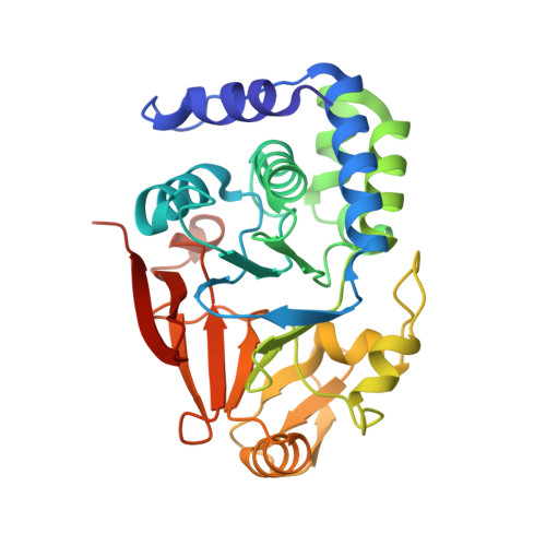
Structural basis for protein phosphatase 1 recruitment by glycogen-targeting subunits

Yu, J.Deng, T.Xiang, S.

(2018) FEBS J 285: 4646-4659

  • DOI: https://doi.org/10.1111/febs.14699
  • Primary Citation of Related Structures:  
    5ZQV, 5ZT0

  • PubMed Abstract: 

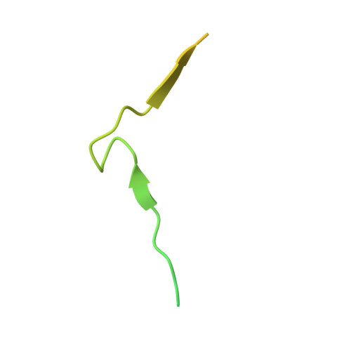
    The rate-limiting enzymes in glycogen metabolism are subject to regulation by reversible phosphorylation. The glycogen-targeted protein phosphatase 1 (PP1) holoenzyme catalyzes their dephosphorylation. It is composed of a catalytic subunit (PP1C) and a glycogen-targeting subunit (G subunit). To date, seven G subunits have been identified. They all contain an RVxF PP1C-binding motif. The interactions between this motif in the skeletal muscle-specific G M and PP1C have been revealed by structural studies. However, whether elements outside of this motif contribute to the interaction with PP1C is not clear. In this study, we found that residues next to the RVxF motif in G M also mediate interactions to PP1C and revealed the mechanism of the interaction by structural studies. Sequence analysis revealed that the PP1C-binding region in G M is highly conserved among G subunits. Consistently, we found that the equivalent region in the liver-enriched G L adopts a similar structure upon binding PP1C. Dephosphorylation experiments indicated that this region and the glycogen-binding region in G M cooperate to stimulate PP1C's activity toward glycogen-associated substrates. DATABASES: The structure factors and coordinates for the PP1Cα-G M (1-99) and PP1Cα-G L (31-105) complexes have been deposited into the Protein Data Bank (http://www.pdb.org), with the accession codes 5ZQV and 5ZT0, respectively.


  • Organizational Affiliation

    CAS Key Laboratory of Nutrition, Metabolism and Food Safety, Shanghai Institute of Nutrition and Health, Shanghai Institutes for Biological Sciences, University of Chinese Academy of Sciences, Chinese Academy of Sciences, Shanghai, China.


Macromolecules
Find similar proteins by:  (by identity cutoff)  |  3D Structure
Entity ID: 1
MoleculeChains Sequence LengthOrganismDetailsImage
Serine/threonine-protein phosphatase PP1-alpha catalytic subunit
A, B, C, D, E
A, B, C, D, E, F
294Mus musculusMutation(s): 0 
Gene Names: Ppp1caPpp1a
EC: 3.1.3.16
UniProt
Find proteins for P62137 (Mus musculus)
Explore P62137 
Go to UniProtKB:  P62137
Entity Groups  
Sequence Clusters30% Identity50% Identity70% Identity90% Identity95% Identity100% Identity
UniProt GroupP62137
Sequence Annotations
Expand
  • Reference Sequence
Find similar proteins by:  (by identity cutoff)  |  3D Structure
Entity ID: 2
MoleculeChains Sequence LengthOrganismDetailsImage
Protein phosphatase 1 regulatory subunit 3B
G, H, I, J
75Homo sapiensMutation(s): 0 
Gene Names: PPP1R3B
UniProt & NIH Common Fund Data Resources
Find proteins for Q86XI6 (Homo sapiens)
Explore Q86XI6 
Go to UniProtKB:  Q86XI6
PHAROS:  Q86XI6
GTEx:  ENSG00000173281 
Entity Groups  
Sequence Clusters30% Identity50% Identity70% Identity90% Identity95% Identity100% Identity
UniProt GroupQ86XI6
Sequence Annotations
Expand
  • Reference Sequence
Small Molecules
Ligands 2 Unique
IDChains Name / Formula / InChI Key2D Diagram3D Interactions
PO4 (Subject of Investigation/LOI)
Query on PO4

Download Ideal Coordinates CCD File 
BA [auth F]
M [auth A]
P [auth B]
S [auth C]
V [auth D]
BA [auth F],
M [auth A],
P [auth B],
S [auth C],
V [auth D],
Y [auth E]
PHOSPHATE ION
O4 P
NBIIXXVUZAFLBC-UHFFFAOYSA-K
MN (Subject of Investigation/LOI)
Query on MN

Download Ideal Coordinates CCD File 
AA [auth F]
K [auth A]
L [auth A]
N [auth B]
O [auth B]
AA [auth F],
K [auth A],
L [auth A],
N [auth B],
O [auth B],
Q [auth C],
R [auth C],
T [auth D],
U [auth D],
W [auth E],
X [auth E],
Z [auth F]
MANGANESE (II) ION
Mn
WAEMQWOKJMHJLA-UHFFFAOYSA-N
Experimental Data & Validation

Experimental Data

  • Method: X-RAY DIFFRACTION
  • Resolution: 3.32 Å
  • R-Value Free: 0.233 
  • R-Value Work: 0.200 
  • R-Value Observed: 0.202 
  • Space Group: P 32
Unit Cell:
Length ( Å )Angle ( ˚ )
a = 106.53α = 90
b = 106.53β = 90
c = 187.51γ = 120
Software Package:
Software NamePurpose
REFMACrefinement
PHENIXrefinement
SCALAdata scaling
iMOSFLMdata reduction
MOLREPphasing

Structure Validation

View Full Validation Report


Ligand Structure Quality Assessment 


Entry History & Funding Information

Deposition Data


Funding OrganizationLocationGrant Number
Ministry of Science and Technology (China)China2011CB910500

Revision History  (Full details and data files)

  • Version 1.0: 2019-03-13
    Type: Initial release
  • Version 1.1: 2023-11-22
    Changes: Data collection, Database references, Derived calculations, Refinement description