5ZI3

MDH3 wild type, apo-form


Experimental Data Snapshot

  • Method: X-RAY DIFFRACTION
  • Resolution: 2.10 Å
  • R-Value Free: 0.265 
  • R-Value Work: 0.202 
  • R-Value Observed: 0.205 

wwPDB Validation   3D Report Full Report


This is version 1.1 of the entry. See complete history


Literature

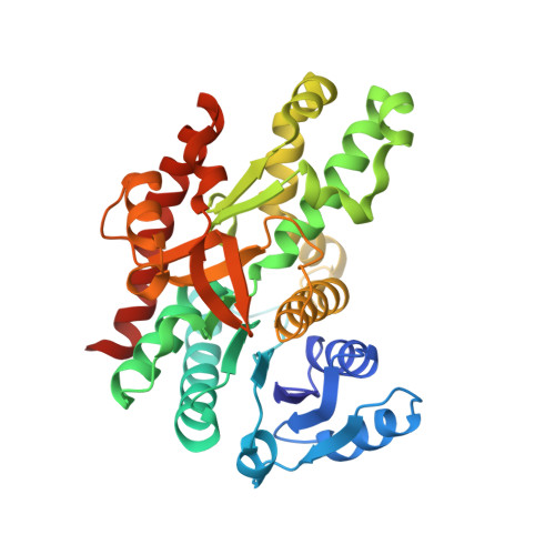
Structure of glyoxysomal malate dehydrogenase (MDH3) from Saccharomyces cerevisiae.

Moriyama, S.Nishio, K.Mizushima, T.

(2018) Acta Crystallogr F Struct Biol Commun 74: 617-624

  • DOI: https://doi.org/10.1107/S2053230X18011895
  • Primary Citation of Related Structures:  
    5ZI2, 5ZI3, 5ZI4

  • PubMed Abstract: 

    Malate dehydrogenase (MDH), a carbohydrate and energy metabolism enzyme in eukaryotes, catalyzes the interconversion of malate to oxaloacetate (OAA) in conjunction with that of nicotinamide adenine dinucleotide (NAD + ) to NADH. Three isozymes of MDH have been reported in Saccharomyces cerevisiae: MDH1, MDH2 and MDH3. MDH1 is a mitochondrial enzyme and a member of the tricarboxylic acid cycle, whereas MDH2 is a cytosolic enzyme that functions in the glyoxylate cycle. MDH3 is a glyoxysomal enzyme that is involved in the reoxidation of NADH, which is produced during fatty-acid β-oxidation. The affinity of MDH3 for OAA is lower than those of MDH1 and MDH2. Here, the crystal structures of yeast apo MDH3, the MDH3-NAD + complex and the MDH3-NAD + -OAA ternary complex were determined. The structure of the ternary complex suggests that the active-site loop is in the open conformation, differing from the closed conformations in mitochondrial and cytosolic malate dehydrogenases.


  • Organizational Affiliation

    Picobiology Institute, Graduate School of Life Science, University of Hyogo, 3-2-1 Kouto, Kamigori-cho, Ako-gun, Hyogo 678-1297, Japan.


Macromolecules
Find similar proteins by:  (by identity cutoff)  |  3D Structure
Entity ID: 1
MoleculeChains Sequence LengthOrganismDetailsImage
Malate dehydrogenase
A, B
345Saccharomyces cerevisiaeMutation(s): 0 
Gene Names: MDH3SCKG_0552
EC: 1.1.1.37
UniProt
Find proteins for P32419 (Saccharomyces cerevisiae (strain ATCC 204508 / S288c))
Explore P32419 
Go to UniProtKB:  P32419
Entity Groups  
Sequence Clusters30% Identity50% Identity70% Identity90% Identity95% Identity100% Identity
UniProt GroupP32419
Sequence Annotations
Expand
  • Reference Sequence
Experimental Data & Validation

Experimental Data

  • Method: X-RAY DIFFRACTION
  • Resolution: 2.10 Å
  • R-Value Free: 0.265 
  • R-Value Work: 0.202 
  • R-Value Observed: 0.205 
  • Space Group: P 21 21 21
Unit Cell:
Length ( Å )Angle ( ˚ )
a = 57.455α = 90
b = 86.645β = 90
c = 132.931γ = 90
Software Package:
Software NamePurpose
REFMACrefinement
HKL-2000data reduction
HKL-2000data scaling
PHASERphasing

Structure Validation

View Full Validation Report


Entry History 

Deposition Data

Revision History  (Full details and data files)

  • Version 1.0: 2018-10-24
    Type: Initial release
  • Version 1.1: 2023-11-22
    Changes: Data collection, Database references, Refinement description