5ZG9

Crystal structure of MoSub1-ssDNA complex in phosphate buffer


Experimental Data Snapshot

  • Method: X-RAY DIFFRACTION
  • Resolution: 2.04 Å
  • R-Value Free: 0.224 
  • R-Value Work: 0.176 
  • R-Value Observed: 0.179 

wwPDB Validation   3D Report Full Report


Ligand Structure Quality Assessment 


This is version 1.1 of the entry. See complete history


Literature

The effect of phosphate ion on the ssDNA binding mode of MoSub1, a Sub1/PC4 homolog from rice blast fungus.

Zhao, Y.Zhang, Y.Huang, J.Wang, S.Yi, L.Zhang, X.Xu, M.Fang, X.Liu, J.

(2019) Proteins 87: 257-264

  • DOI: https://doi.org/10.1002/prot.25647
  • Primary Citation of Related Structures:  
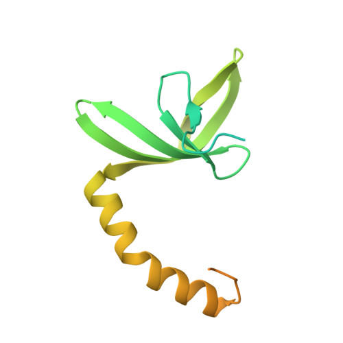
    5ZG9

  • PubMed Abstract: 

    MoSub1 is an ortholog of yeast single stranded DNA binding protein Sub1 or human PC4 from rice blast fungus. All of them share a similar DNA binding region and may have similar biological roles. The well-studied Sub1/PC4 has been reported to play multiple roles in DNA metabolic processes, such as transcription and DNA repair and their DNA binding capacity is significantly affected by phosphorylation. Here, we determined the crystal structure of MoSub1 complexed with ssDNA in a phosphate solution. The crystal structure of the MoSub1-ssDNA complex was solved to a resolution of 2.04 Å. A phosphate ion at the interface of the protein-DNA interaction of the complex bridged the lys84 of the protein and two nucleotides. The DNA was bound in novel mode (L mode) in the MoSub1 complex in the presence of phosphate ions, while DNA bound in the straight mode in the absence of the phosphate ion and in U mode in the same binding motif of the PC4-ssDNA complex. The crystal structure of the complex and a small-angle X-ray scattering analysis revealed that the phosphate ion at the protein-DNA interface affected the DNA binding mode of MoSub1 to oligo-DNA and provided a new structural clue for studying its functions.


  • Organizational Affiliation

    Department of Plant Pathology, and MOA Key Laboratory of Plant Pathology, China Agricultural University, Beijing, China.


Macromolecules

Find similar proteins by:  (by identity cutoff)  |  3D Structure
Entity ID: 1
MoleculeChains Sequence LengthOrganismDetailsImage
MoSub1
A, B
169Pyricularia oryzae P131Mutation(s): 0 
Gene Names: OOW_P131scaffold01307g15
UniProt
Find proteins for L7IX95 (Pyricularia oryzae (strain P131))
Explore L7IX95 
Go to UniProtKB:  L7IX95
Entity Groups  
Sequence Clusters30% Identity50% Identity70% Identity90% Identity95% Identity100% Identity
UniProt GroupL7IX95
Sequence Annotations
Expand
  • Reference Sequence

Find similar nucleic acids by:  Sequence   |   3D Structure  

Entity ID: 2
MoleculeChains LengthOrganismImage
DNA (5'-D(P*TP*TP*TP*TP*TP*TP*TP*TP*TP*TP*TP*TP*TP*TP*TP*TP*TP*TP*TP*G)-3')20synthetic construct
Sequence Annotations
Expand
  • Reference Sequence
Experimental Data & Validation

Experimental Data

  • Method: X-RAY DIFFRACTION
  • Resolution: 2.04 Å
  • R-Value Free: 0.224 
  • R-Value Work: 0.176 
  • R-Value Observed: 0.179 
  • Space Group: I 1 2 1
Unit Cell:
Length ( Å )Angle ( ˚ )
a = 42.01α = 90
b = 80.19β = 91.88
c = 76.91γ = 90
Software Package:
Software NamePurpose
PHENIXrefinement
XDSdata reduction
XDSdata scaling
PHENIXphasing

Structure Validation

View Full Validation Report


Ligand Structure Quality Assessment 


Entry History & Funding Information

Deposition Data


Funding OrganizationLocationGrant Number
Ministry of Science and Technology (China)China2017YFD0200500
Ministry of Science and Technology (China)China2017YFD0201100

Revision History  (Full details and data files)

  • Version 1.0: 2019-03-27
    Type: Initial release
  • Version 1.1: 2024-03-27
    Changes: Data collection, Database references