5YS4

Crystal structure of retroviral protease-like domain of Ddi1 from Leishmania major


Experimental Data Snapshot

  • Method: X-RAY DIFFRACTION
  • Resolution: 2.30 Å
  • R-Value Free: 0.259 
  • R-Value Work: 0.236 
  • R-Value Observed: 0.238 

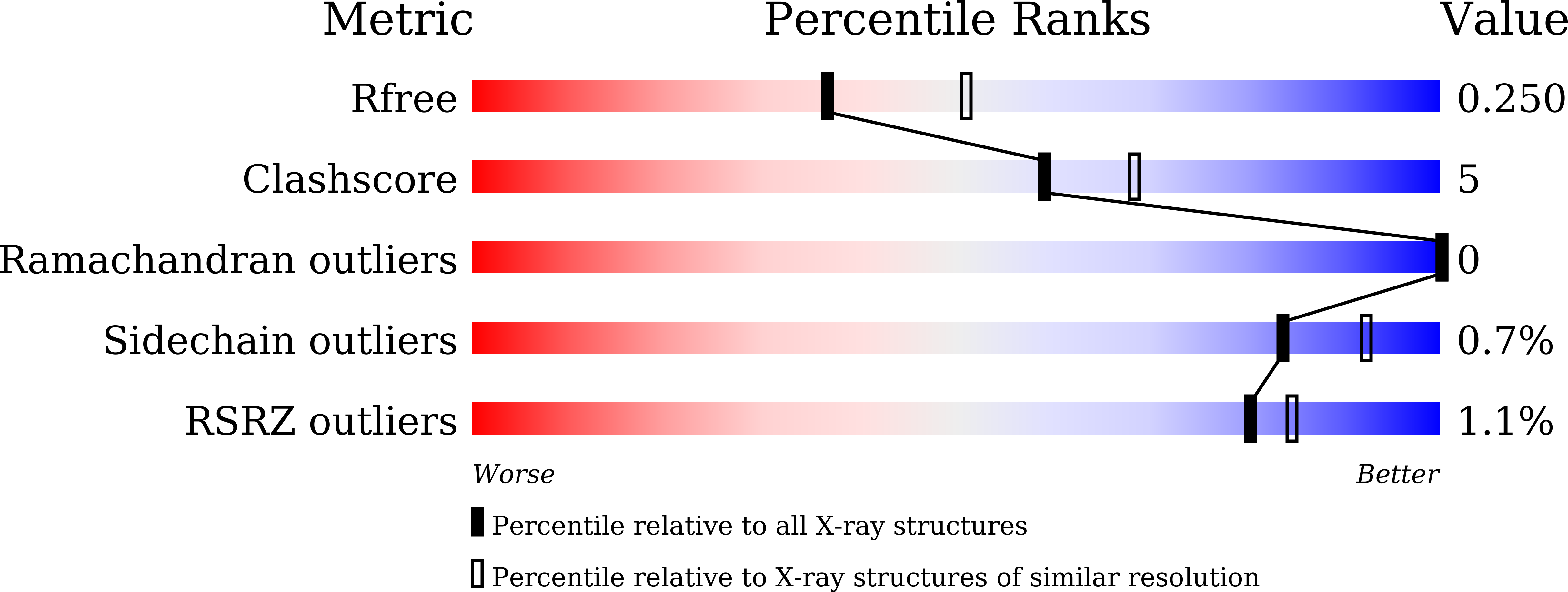
wwPDB Validation   3D Report Full Report


This is version 1.1 of the entry. See complete history


Literature

Crystal structure of the retroviral protease-like domain of a protozoal DNA damage-inducible 1 protein.

Kumar, S.Suguna, K.

(2018) FEBS Open Bio 8: 1379-1394

  • DOI: https://doi.org/10.1002/2211-5463.12491
  • Primary Citation of Related Structures:  
    5YQ8, 5YS4

  • PubMed Abstract: 

    DNA damage-inducible 1 (Ddi1) is a multidomain protein with one of the domains being retropepsin-like. HIV-1 protease inhibitors were found to reduce opportunistic infections caused by pathogens like Leishmania and Plasmodium , and some of them were shown to inhibit the growth of these parasites. In Leishmania , Ddi1 was identified as a likely target of the inhibitors. We report the crystal structure of the retropepsin-like domain of Ddi1 from Leishmania major as a dimer with clear density for the critical 'flap' region. We have characterized binding with one of the HIV-1 protease inhibitors in solution using bio-layer interferometry and by docking. Further, we have performed molecular dynamics (MD) simulation studies that show that the protein undergoes a conformational change from open to semi-open and closed forms with the closing of the flexible flap over the active site.


  • Organizational Affiliation

    Molecular Biophysics Unit Indian Institute of Science Bangalore India.


Macromolecules
Find similar proteins by:  (by identity cutoff)  |  3D Structure
Entity ID: 1
MoleculeChains Sequence LengthOrganismDetailsImage
DNA-damage inducible protein DDI1-like protein
A, B, C, D, E
A, B, C, D, E, F
130Leishmania majorMutation(s): 0 
Gene Names: LMJF_01_0610
UniProt
Find proteins for I7HUG0 (Leishmania major)
Explore I7HUG0 
Go to UniProtKB:  I7HUG0
Entity Groups  
Sequence Clusters30% Identity50% Identity70% Identity90% Identity95% Identity100% Identity
UniProt GroupI7HUG0
Sequence Annotations
Expand
  • Reference Sequence
Experimental Data & Validation

Experimental Data

  • Method: X-RAY DIFFRACTION
  • Resolution: 2.30 Å
  • R-Value Free: 0.259 
  • R-Value Work: 0.236 
  • R-Value Observed: 0.238 
  • Space Group: P 1
Unit Cell:
Length ( Å )Angle ( ˚ )
a = 43.79α = 76.56
b = 56.78β = 84.48
c = 91.4γ = 89.99
Software Package:
Software NamePurpose
PHENIXrefinement
PDB_EXTRACTdata extraction
iMOSFLMdata reduction
Aimlessdata scaling
PHASERphasing

Structure Validation

View Full Validation Report


Entry History & Funding Information

Deposition Data


Funding OrganizationLocationGrant Number
Other governmentIndiaBT/PR1770/BRB/10/1505/2016

Revision History  (Full details and data files)

  • Version 1.0: 2018-11-21
    Type: Initial release
  • Version 1.1: 2023-11-22
    Changes: Data collection, Database references, Refinement description