Search of multiple hot spots on the surface of peptidyl-tRNA hydrolase: structural, binding and antibacterial studies.
Kaushik, S., Iqbal, N., Singh, N., Sikarwar, J.S., Singh, P.K., Sharma, P., Kaur, P., Sharma, S., Owais, M., Singh, T.P.(2018) Biochem J 475: 547-560
- PubMed: 29301982
- DOI: https://doi.org/10.1042/BCJ20170666
- Primary Citation of Related Structures:
5Y98, 5Y9A - PubMed Abstract:
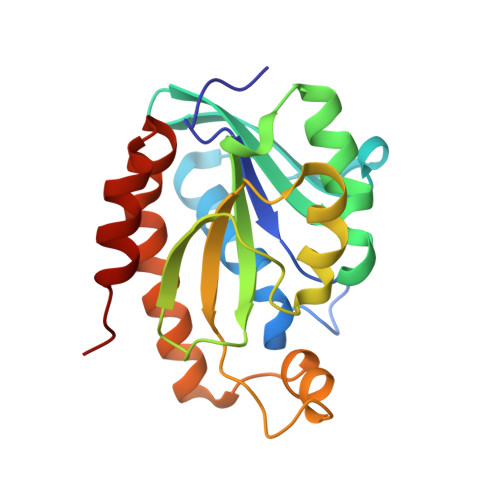
Peptidyl-tRNA hydrolase (Pth) catalyzes the breakdown of peptidyl-tRNA into peptide and tRNA components. Pth from Acinetobacter baumannii ( Ab Pth) was cloned, expressed, purified and crystallized in a native unbound ( Ab Pth-N) state and in a bound state with the phosphate ion and cytosine arabinoside (cytarabine) ( Ab Pth-C). Structures of Ab Pth-N and Ab Pth-C were determined at 1.36 and 1.10 Å resolutions, respectively. The structure of Ab Pth-N showed that the active site is filled with water molecules. In the structure of Ab Pth-C, a phosphate ion is present in the active site, while cytarabine is bound in a cleft which is located away from the catalytic site. The cytarabine-binding site is formed with residues: Gln19, Trp27, Glu30, Gln31, Lys152, Gln158 and Asp162. In the structure of Ab Pth-N, the side chains of two active-site residues, Asn70 and Asn116, were observed in two conformations. Upon binding of the phosphate ion in the active site, the side chains of both residues were ordered to single conformations. Since Trp27 is present at the cytarabine-binding site, the fluorescence studies were carried out which gave a dissociation constant ( K D ) of 3.3 ± 0.8 × 10 -7 M for cytarabine. The binding studies using surface plasmon resonance gave a K D value of 3.7 ± 0.7 × 10 -7 M. The bacterial inhibition studies using the agar diffusion method and the biofilm inhibition assay established the strong antimicrobial potential of cytarabine. It also indicated that cytarabine inhibited Gram-negative bacteria more profoundly when compared with Gram-positive bacteria in a dose-dependent manner. Cytarabine was also effective against the drug-resistant bacteria both alone as well as in combination with other antibiotics.
Organizational Affiliation:
Department of Biophysics, All India Institute of Medical Sciences, Ansari Nagar, New Delhi 110 029, India.