5XWS

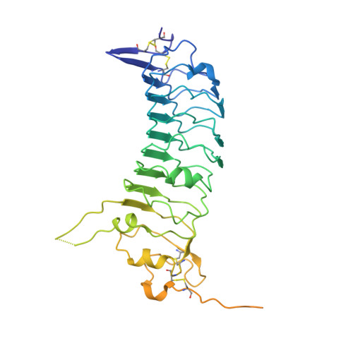
Crystal structure of SALM5 LRR-Ig


Experimental Data Snapshot

  • Method: X-RAY DIFFRACTION
  • Resolution: 3.08 Å
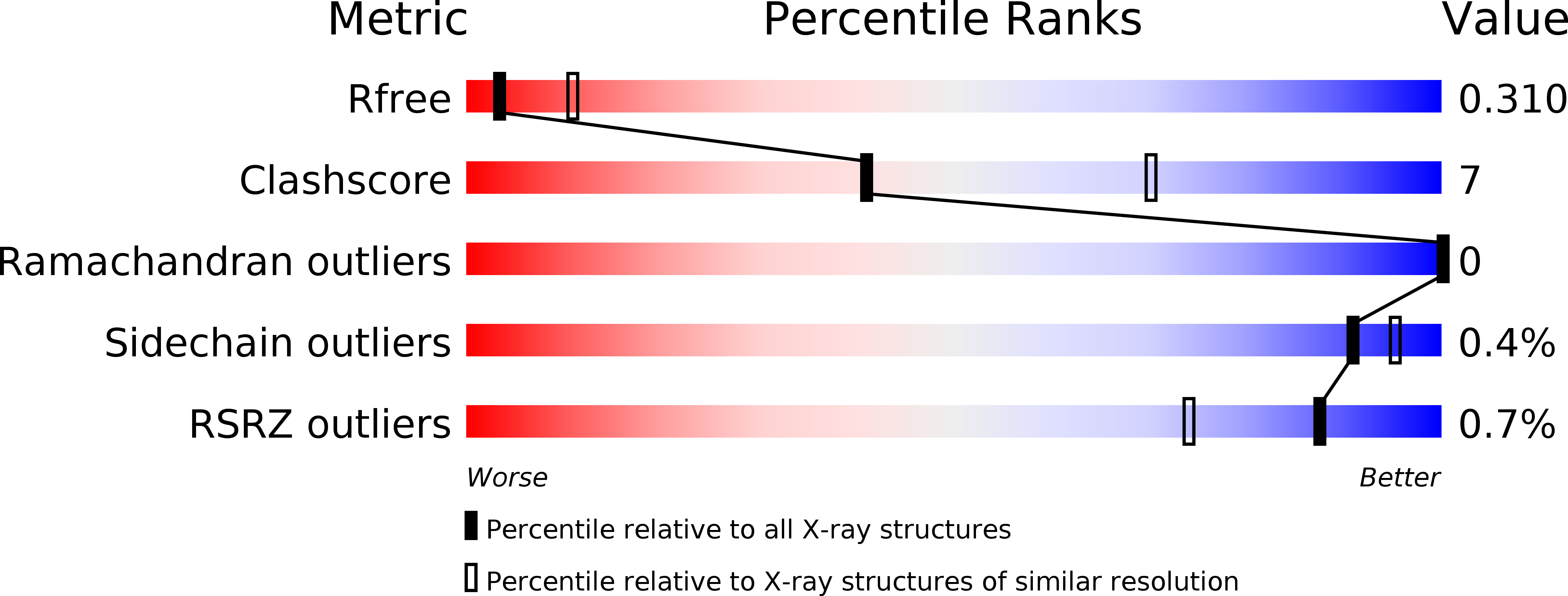
  • R-Value Free: 0.308 
  • R-Value Work: 0.268 
  • R-Value Observed: 0.270 

wwPDB Validation   3D Report Full Report


This is version 2.0 of the entry. See complete history


Literature

Structural basis of trans-synaptic interactions between PTP delta and SALMs for inducing synapse formation.

Goto-Ito, S.Yamagata, A.Sato, Y.Uemura, T.Shiroshima, T.Maeda, A.Imai, A.Mori, H.Yoshida, T.Fukai, S.

(2018) Nat Commun 9: 269-269

  • DOI: https://doi.org/10.1038/s41467-017-02417-z
  • Primary Citation of Related Structures:  
    5XWS, 5XWT, 5XWU

  • PubMed Abstract: 

    Synapse formation is triggered by trans-synaptic interactions of cell adhesion molecules, termed synaptic organizers. Three members of type-II receptor protein tyrosine phosphatases (classified as type-IIa RPTPs; PTPδ, PTPσ and LAR) are known as presynaptic organizers. Synaptic adhesion-like molecules (SALMs) have recently emerged as a family of postsynaptic organizers. Although all five SALM isoforms can bind to the type-IIa RPTPs, only SALM3 and SALM5 reportedly have synaptogenic activities depending on their binding. Here, we report the crystal structures of apo-SALM5, and PTPδ-SALM2 and PTPδ-SALM5 complexes. The leucine-rich repeat (LRR) domains of SALMs interact with the second immunoglobulin-like (Ig) domain of PTPδ, whereas the Ig domains of SALMs interact with both the second and third Ig domains of PTPδ. Unexpectedly, the structures exhibit the LRR-mediated 2:2 complex. Our synaptogenic co-culture assay using site-directed SALM5 mutants demonstrates that presynaptic differentiation induced by PTPδ-SALM5 requires the dimeric property of SALM5.


  • Organizational Affiliation

    Institute of Molecular and Cellular Biosciences, The University of Tokyo, Tokyo, 113-0032, Japan.


Macromolecules
Find similar proteins by:  (by identity cutoff)  |  3D Structure
Entity ID: 1
MoleculeChains Sequence LengthOrganismDetailsImage
Leucine-rich repeat and fibronectin type-III domain-containing protein 5367Homo sapiensMutation(s): 0 
Gene Names: LRFN5C14orf146SALM5
UniProt & NIH Common Fund Data Resources
Find proteins for Q96NI6 (Homo sapiens)
Explore Q96NI6 
Go to UniProtKB:  Q96NI6
PHAROS:  Q96NI6
GTEx:  ENSG00000165379 
Entity Groups  
Sequence Clusters30% Identity50% Identity70% Identity90% Identity95% Identity100% Identity
UniProt GroupQ96NI6
Sequence Annotations
Expand
  • Reference Sequence
Oligosaccharides

Help

Entity ID: 2
MoleculeChains Length2D Diagram Glycosylation3D Interactions
2-acetamido-2-deoxy-beta-D-glucopyranose-(1-4)-2-acetamido-2-deoxy-beta-D-glucopyranose
B
2N-Glycosylation
Glycosylation Resources
GlyTouCan:  G42666HT
GlyCosmos:  G42666HT
GlyGen:  G42666HT
Experimental Data & Validation

Experimental Data

  • Method: X-RAY DIFFRACTION
  • Resolution: 3.08 Å
  • R-Value Free: 0.308 
  • R-Value Work: 0.268 
  • R-Value Observed: 0.270 
  • Space Group: P 63 2 2
Unit Cell:
Length ( Å )Angle ( ˚ )
a = 154.888α = 90
b = 154.888β = 90
c = 91.262γ = 120
Software Package:
Software NamePurpose
PHENIXrefinement
HKL-2000data reduction
HKL-2000data scaling
MOLREPphasing

Structure Validation

View Full Validation Report


Entry History & Funding Information

Deposition Data


Funding OrganizationLocationGrant Number
JST CRESTJapan--
MEXT/JSPSJapan22121003
MEXT/JSPSJapan16H04749

Revision History  (Full details and data files)

  • Version 1.0: 2018-06-06
    Type: Initial release
  • Version 2.0: 2020-07-29
    Type: Remediation
    Reason: Carbohydrate remediation
    Changes: Atomic model, Data collection, Derived calculations, Structure summary